6WWZ

Cryo-EM structure of the human chemokine receptor CCR6 in complex with CCL20 and a Go protein


Experimental Data Snapshot

  • Method: ELECTRON MICROSCOPY
  • Resolution: 3.34 Å
  • Aggregation State: PARTICLE 
  • Reconstruction Method: SINGLE PARTICLE 

wwPDB Validation   3D Report Full Report


This is version 1.1 of the entry. See complete history


Literature

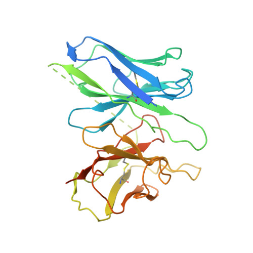
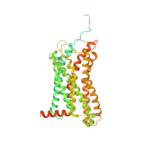
Structural basis for chemokine receptor CCR6 activation by the endogenous protein ligand CCL20.

Wasilko, D.J.Johnson, Z.L.Ammirati, M.Che, Y.Griffor, M.C.Han, S.Wu, H.

(2020) Nat Commun 11: 3031-3031

  • DOI: https://doi.org/10.1038/s41467-020-16820-6
  • Primary Citation of Related Structures:  
    6WWZ

  • PubMed Abstract: 

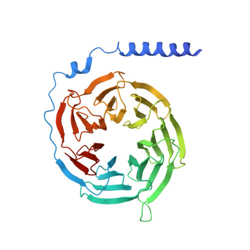
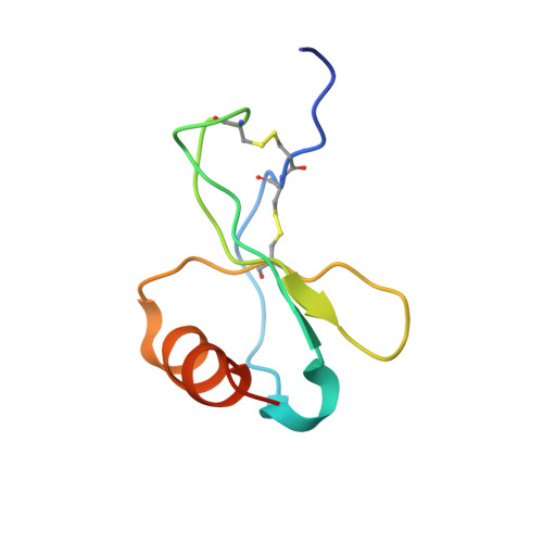
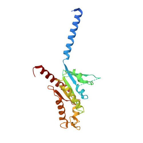
    Chemokines are important protein-signaling molecules that regulate various immune responses by activating chemokine receptors which belong to the G protein-coupled receptor (GPCR) superfamily. Despite the substantial progression of our structural understanding of GPCR activation by small molecule and peptide agonists, the molecular mechanism of GPCR activation by protein agonists remains unclear. Here, we present a 3.3-Å cryo-electron microscopy structure of the human chemokine receptor CCR6 bound to its endogenous ligand CCL20 and an engineered Go. CCL20 binds in a shallow extracellular pocket, making limited contact with the core 7-transmembrane (TM) bundle. The structure suggests that this mode of binding induces allosterically a rearrangement of a noncanonical toggle switch and the opening of the intracellular crevice for G protein coupling. Our results demonstrate that GPCR activation by a protein agonist does not always require substantial interactions between ligand and the 7TM core region.


  • Organizational Affiliation

    Discovery Sciences, Medicine Design, Pfizer Worldwide Research and Development, Groton, CT, 06340, USA.


Macromolecules
Find similar proteins by:  (by identity cutoff)  |  3D Structure
Entity ID: 1
MoleculeChains Sequence LengthOrganismDetailsImage
Guanine nucleotide-binding protein G(I)/G(S)/G(T) subunit beta-1A [auth B]351Rattus norvegicusMutation(s): 0 
Gene Names: Gnb1
UniProt
Find proteins for P54311 (Rattus norvegicus)
Explore P54311 
Go to UniProtKB:  P54311
Entity Groups  
Sequence Clusters30% Identity50% Identity70% Identity90% Identity95% Identity100% Identity
UniProt GroupP54311
Sequence Annotations
Expand
  • Reference Sequence
Find similar proteins by:  (by identity cutoff)  |  3D Structure
Entity ID: 2
MoleculeChains Sequence LengthOrganismDetailsImage
C-C motif chemokine 20B [auth C]70Homo sapiensMutation(s): 0 
Gene Names: CCL20LARCMIP3ASCYA20
Membrane Entity: Yes 
UniProt & NIH Common Fund Data Resources
Find proteins for P78556 (Homo sapiens)
Explore P78556 
Go to UniProtKB:  P78556
PHAROS:  P78556
GTEx:  ENSG00000115009 
Entity Groups  
Sequence Clusters30% Identity50% Identity70% Identity90% Identity95% Identity100% Identity
UniProt GroupP78556
Sequence Annotations
Expand
  • Reference Sequence
Find similar proteins by:  (by identity cutoff)  |  3D Structure
Entity ID: 3
MoleculeChains Sequence LengthOrganismDetailsImage
Guanine nucleotide-binding protein G(I)/G(S)/G(O) subunit gamma-2C [auth Y]68Rattus norvegicusMutation(s): 0 
Membrane Entity: Yes 
UniProt & NIH Common Fund Data Resources
Find proteins for P59768 (Homo sapiens)
Explore P59768 
Go to UniProtKB:  P59768
PHAROS:  P59768
GTEx:  ENSG00000186469 
Entity Groups  
Sequence Clusters30% Identity50% Identity70% Identity90% Identity95% Identity100% Identity
UniProt GroupP59768
Sequence Annotations
Expand
  • Reference Sequence
Find similar proteins by:  (by identity cutoff)  |  3D Structure
Entity ID: 4
MoleculeChains Sequence LengthOrganismDetailsImage
Guanine nucleotide-binding protein G(o) subunit alpha,Guanine nucleotide-binding protein G(o) subunit alphaD [auth A]250Homo sapiensMutation(s): 0 
Gene Names: GNAO1
Membrane Entity: Yes 
UniProt & NIH Common Fund Data Resources
Find proteins for P09471 (Homo sapiens)
Explore P09471 
Go to UniProtKB:  P09471
PHAROS:  P09471
GTEx:  ENSG00000087258 
Entity Groups  
Sequence Clusters30% Identity50% Identity70% Identity90% Identity95% Identity100% Identity
UniProt GroupP09471
Sequence Annotations
Expand
  • Reference Sequence
Find similar proteins by:  (by identity cutoff)  |  3D Structure
Entity ID: 5
MoleculeChains Sequence LengthOrganismDetailsImage
scFv16E [auth S]287Mus musculusMutation(s): 0 
Entity Groups  
Sequence Clusters30% Identity50% Identity70% Identity90% Identity95% Identity100% Identity
Sequence Annotations
Expand
  • Reference Sequence
Find similar proteins by:  (by identity cutoff)  |  3D Structure
Entity ID: 6
MoleculeChains Sequence LengthOrganismDetailsImage
C-C chemokine receptor type 6,C-C chemokine receptor type 6F [auth R]529Homo sapiensMutation(s): 0 
Membrane Entity: Yes 
UniProt & NIH Common Fund Data Resources
Find proteins for P51684 (Homo sapiens)
Explore P51684 
Go to UniProtKB:  P51684
PHAROS:  P51684
GTEx:  ENSG00000112486 
Find proteins for P0ABE7 (Escherichia coli)
Explore P0ABE7 
Go to UniProtKB:  P0ABE7
Entity Groups  
Sequence Clusters30% Identity50% Identity70% Identity90% Identity95% Identity100% Identity
UniProt GroupsP0ABE7P51684
Sequence Annotations
Expand
  • Reference Sequence
Experimental Data & Validation

Experimental Data

  • Method: ELECTRON MICROSCOPY
  • Resolution: 3.34 Å
  • Aggregation State: PARTICLE 
  • Reconstruction Method: SINGLE PARTICLE 
EM Software:
TaskSoftware PackageVersion
RECONSTRUCTIONRELION3.0.5
MODEL REFINEMENTCoot0.8.9.2
MODEL REFINEMENTPHENIXdev-3026
MODEL REFINEMENTREFMACCCP4 7.0
MODEL REFINEMENTCoot0.8.9.2
MODEL REFINEMENTPHENIXdev-3026
MODEL REFINEMENTREFMACCCP4 7.0

Structure Validation

View Full Validation Report


Entry History 

Deposition Data

Revision History  (Full details and data files)

  • Version 1.0: 2020-06-24
    Type: Initial release
  • Version 1.1: 2020-07-01
    Changes: Database references