6W9Q

Peptide-bound SARS-CoV-2 Nsp9 RNA-replicase


Experimental Data Snapshot

  • Method: X-RAY DIFFRACTION
  • Resolution: 2.05 Å
  • R-Value Free: 0.246 
  • R-Value Work: 0.233 
  • R-Value Observed: 0.233 

wwPDB Validation   3D Report Full Report


This is version 1.3 of the entry. See complete history


Literature

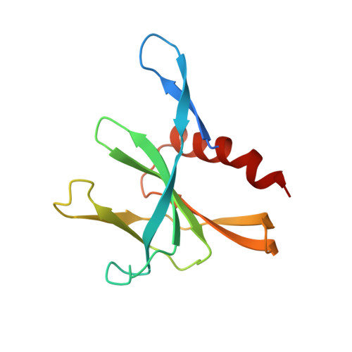
Crystal Structure of the SARS-CoV-2 Non-structural Protein 9, Nsp9.

Littler, D.R.Gully, B.S.Colson, R.N.Rossjohn, J.

(2020) iScience 23: 101258-101258

  • DOI: https://doi.org/10.1016/j.isci.2020.101258
  • Primary Citation of Related Structures:  
    6W9Q, 6WXD

  • PubMed Abstract: 

    Many of the SARS-CoV-2 proteins have related counterparts across the Severe Acute Respiratory Syndrome (SARS-CoV) family. One such protein is non-structural protein 9 (Nsp9), which is thought to mediate viral replication, overall virulence, and viral genomic RNA reproduction. We sought to better characterize the SARS-CoV-2 Nsp9 and subsequently solved its X-ray crystal structure, in an apo form and, unexpectedly, in a peptide-bound form with a sequence originating from a rhinoviral 3C protease sequence (LEVL). The SARS-CoV-2 Nsp9 structure revealed the high level of structural conservation within the Nsp9 family. The exogenous peptide binding site is close to the dimer interface and impacted the relative juxtapositioning of the monomers within the homodimer. We have established a protocol for the production of SARS-CoV-2 Nsp9, determined its structure, and identified a peptide-binding site that warrants further study to understanding Nsp9 function.


  • Organizational Affiliation

    Infection and Immunity Program, Department of Biochemistry and Molecular Biology, Biomedicine Discovery Institute, Monash University, Clayton, VIC, Australia; Australian Research Council Centre of Excellence for Advanced Molecular Imaging, Monash University, Clayton, VIC, Australia. Electronic address: dene.littler@monash.edu.


Macromolecules
Find similar proteins by:  (by identity cutoff)  |  3D Structure
Entity ID: 1
MoleculeChains Sequence LengthOrganismDetailsImage
3C-like proteinase peptide, Non-structural protein 9 fusion133Severe acute respiratory syndrome coronavirus 2Mutation(s): 0 
Gene Names: rep1a-1b
UniProt
Find proteins for P0DTD1 (Severe acute respiratory syndrome coronavirus 2)
Explore P0DTD1 
Go to UniProtKB:  P0DTD1
Entity Groups  
Sequence Clusters30% Identity50% Identity70% Identity90% Identity95% Identity100% Identity
UniProt GroupP0DTD1
Sequence Annotations
Expand
  • Reference Sequence
Small Molecules
Ligands 1 Unique
IDChains Name / Formula / InChI Key2D Diagram3D Interactions
PO4
Query on PO4

Download Ideal Coordinates CCD File 
B [auth A]PHOSPHATE ION
O4 P
NBIIXXVUZAFLBC-UHFFFAOYSA-K
Experimental Data & Validation

Experimental Data

  • Method: X-RAY DIFFRACTION
  • Resolution: 2.05 Å
  • R-Value Free: 0.246 
  • R-Value Work: 0.233 
  • R-Value Observed: 0.233 
  • Space Group: P 43 2 2
Unit Cell:
Length ( Å )Angle ( ˚ )
a = 59.011α = 90
b = 59.011β = 90
c = 85.649γ = 90
Software Package:
Software NamePurpose
PHENIXrefinement
XDSdata reduction
SCALAdata scaling
PHASERphasing

Structure Validation

View Full Validation Report


Entry History 

Deposition Data

Revision History  (Full details and data files)

  • Version 1.0: 2020-04-08
    Type: Initial release
  • Version 1.1: 2020-05-06
    Changes: Database references, Source and taxonomy, Structure summary
  • Version 1.2: 2020-09-09
    Changes: Database references
  • Version 1.3: 2023-10-18
    Changes: Data collection, Database references, Refinement description