1BDO

STRUCTURE OF THE BIOTINYL DOMAIN OF ACETYL-COENZYME A CARBOXYLASE DETERMINED BY MAD PHASING


Experimental Data Snapshot

  • Method: X-RAY DIFFRACTION
  • Resolution: 1.80 Å
  • R-Value Work: 
    0.189 (Depositor), 0.180 (DCC) 
  • R-Value Observed: 
    0.189 (Depositor) 

wwPDB Validation   3D Report Full Report


Ligand Structure Quality Assessment 

Created with Raphaël 2.3.0Worse 01 BetterLigand structure goodness of fit to experimental dataBest fitted BTNClick on this verticalbar to view details

This is version 1.5 of the entry. See complete history


Literature

Structure of the biotinyl domain of acetyl-coenzyme A carboxylase determined by MAD phasing.

Athappilly, F.K.Hendrickson, W.A.

(1995) Structure 3: 1407-1419

  • DOI: https://doi.org/10.1016/s0969-2126(01)00277-5
  • Primary Citation of Related Structures:  
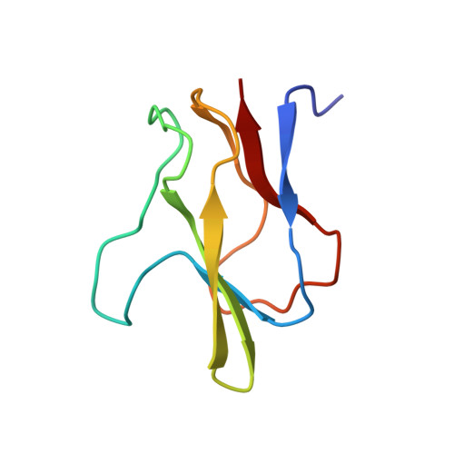
    1BDO

  • PubMed Abstract: 

    Acetyl-coenzyme A carboxylase catalyzes the first committed step of fatty acid biosynthesis. Universally, this reaction involves three functional components all related to a carboxybiotinyl intermediate. A biotinyl domain shuttles its covalently attached biotin prosthetic group between the active sites of a biotin carboxylase and a carboxyl transferase. In Escherichia coli, the three components reside in separate subunits: a biotinyl domain is the functional portion of one of these, biotin carboxy carrier protein (BCCP). We have expressed natural and selenomethionyl (Se-met) BCCP from E. coli as biotinylated recombinant proteins, proteolyzed them with subtilisin Carlsberg to produce the biotinyl domains BCCP and Se-met BCCPsc, determined the crystal structure of Se-met BCCPsc using a modified version of the multiwavelength anomalous diffraction (MAD) phasing protocol, and refined the structure for the natural BCCPsc at 1.8 A resolution. The structure may be described as a capped beta sandwich with quasi-dyad symmetry. Each half contains a characteristic hammerhead motif. The biotinylated lysin is located at a hairpin beta turn which connects the two symmetric halves of the molecule, and its biotinyl group interacts with a non-symmetric protrusion from the core. This first crystal structure of a biotinyl domain helps to unravel the central role of such domains in reactions catalyzed by biotin-dependent carboxylases. The hammerhead structure observed twice in BCCPsc may be regarded as the basic structural motif of biotinyl and lipoyl domains of a superfamily of enzymes. The new MAD phasing techniques developed in the course of determining this structure enhance the power of the MAD method.


  • Organizational Affiliation

    Department of Biochemistry and Molecular Biophysics, College of Physicians and Surgeons of Columbia University, New York, NY 10032, USA.


Macromolecules
Find similar proteins by:  (by identity cutoff)  |  3D Structure
Entity ID: 1
MoleculeChains Sequence LengthOrganismDetailsImage
ACETYL-COA CARBOXYLASE80Escherichia coliMutation(s): 0 
Gene Names: BIOTIN CARBOXYL CARRIER PROTEI
EC: 6.4.1.2
UniProt
Find proteins for P0ABD8 (Escherichia coli (strain K12))
Explore P0ABD8 
Go to UniProtKB:  P0ABD8
Entity Groups  
Sequence Clusters30% Identity50% Identity70% Identity90% Identity95% Identity100% Identity
UniProt GroupP0ABD8
Sequence Annotations
Expand
  • Reference Sequence
Small Molecules
Ligands 1 Unique
IDChains Name / Formula / InChI Key2D Diagram3D Interactions
BTN
Query on BTN

Download Ideal Coordinates CCD File 
B [auth A]BIOTIN
C10 H16 N2 O3 S
YBJHBAHKTGYVGT-ZKWXMUAHSA-N
Experimental Data & Validation

Experimental Data

  • Method: X-RAY DIFFRACTION
  • Resolution: 1.80 Å
  • R-Value Work:  0.189 (Depositor), 0.180 (DCC) 
  • R-Value Observed: 0.189 (Depositor) 
Space Group: P 21 21 2
Unit Cell:
Length ( Å )Angle ( ˚ )
a = 65.46α = 90
b = 37.26β = 90
c = 35.45γ = 90
Software Package:
Software NamePurpose
X-PLORmodel building
X-PLORrefinement
DENZOdata reduction
X-PLORphasing

Structure Validation

View Full Validation Report


Ligand Structure Quality Assessment 

Created with Raphaël 2.3.0Worse 01 BetterLigand structure goodness of fit to experimental dataBest fitted BTNClick on this verticalbar to view details

Entry History 

Deposition Data

Revision History  (Full details and data files)

  • Version 1.0: 1996-08-01
    Type: Initial release
  • Version 1.1: 2008-03-24
    Changes: Version format compliance
  • Version 1.2: 2011-07-13
    Changes: Version format compliance
  • Version 1.3: 2011-11-16
    Changes: Atomic model
  • Version 1.4: 2024-06-05
    Changes: Data collection, Database references, Derived calculations, Other
  • Version 1.5: 2024-10-23
    Changes: Structure summary