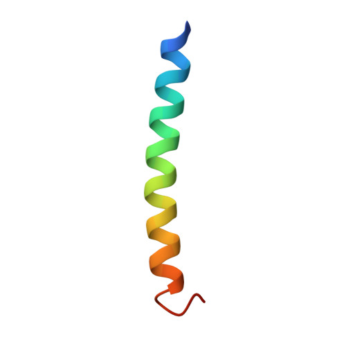
1IJ3

GCN4-pVSL Coiled-coil trimer with Serine at the a(16) position


Experimental Data Snapshot

  • Method: X-RAY DIFFRACTION
  • Resolution: 1.80 Å
  • R-Value Free: 0.269 
  • R-Value Work: 0.220 
  • R-Value Observed: 0.220 

wwPDB Validation   3D Report Full Report


This is version 1.4 of the entry. See complete history


Literature

Buried polar residues in coiled-coil interfaces.

Akey, D.L.Malashkevich, V.N.Kim, P.S.

(2001) Biochemistry 40: 6352-6360

  • DOI: https://doi.org/10.1021/bi002829w
  • Primary Citation of Related Structures:  
    1IJ0, 1IJ1, 1IJ2, 1IJ3

  • PubMed Abstract: 

    Coiled coils, estimated to constitute 3-5% of the encoded residues in most genomes, are characterized by a heptad repeat, (abcdefg)(n), where the buried a and d positions form the interface between multiple alpha-helices. Although generally hydrophobic, a substantial fraction ( approximately 20%) of these a- and d-position residues are polar or charged. We constructed variants of the well-characterized coiled coil GCN4-p1 with a single polar residue (Asn, Gln, Ser, or Thr) at either an a or a d position. The stability and oligomeric specificity of each variant were measured, and crystal structures of coiled-coil trimers with threonine or serine at either an a or a d position were determined. The structures show how single polar residues in the interface affect not only local packing, but also overall coiled-coil geometry as seen by changes in the Crick supercoil parameters and core cavity volumes.


  • Organizational Affiliation

    Howard Hughes Medical Institute, Whitehead Institute for Biomedical Research, Department of Biology, Massachusetts Institute of Technology, Cambridge, Massachusetts 02142, USA.


Macromolecules
Find similar proteins by:  (by identity cutoff)  |  3D Structure
Entity ID: 1
MoleculeChains Sequence LengthOrganismDetailsImage
general control protein GCN4
A, B, C
34N/AMutation(s): 1 
UniProt
Find proteins for P03069 (Saccharomyces cerevisiae (strain ATCC 204508 / S288c))
Explore P03069 
Go to UniProtKB:  P03069
Entity Groups  
Sequence Clusters30% Identity50% Identity70% Identity90% Identity95% Identity100% Identity
UniProt GroupP03069
Sequence Annotations
Expand
  • Reference Sequence
Experimental Data & Validation

Experimental Data

  • Method: X-RAY DIFFRACTION
  • Resolution: 1.80 Å
  • R-Value Free: 0.269 
  • R-Value Work: 0.220 
  • R-Value Observed: 0.220 
  • Space Group: P 1
Unit Cell:
Length ( Å )Angle ( ˚ )
a = 25.564α = 86.47
b = 25.839β = 84.49
c = 34.373γ = 79.4
Software Package:
Software NamePurpose
AMoREphasing
CNSrefinement
DENZOdata reduction
SCALEPACKdata scaling

Structure Validation

View Full Validation Report


Entry History 

Deposition Data

Revision History  (Full details and data files)

  • Version 1.0: 2001-08-08
    Type: Initial release
  • Version 1.1: 2008-04-27
    Changes: Version format compliance
  • Version 1.2: 2011-07-13
    Changes: Version format compliance
  • Version 1.3: 2021-10-27
    Changes: Database references, Derived calculations
  • Version 1.4: 2024-10-16
    Changes: Data collection, Structure summary