1MUJ

Crystal structure of murine class II MHC I-Ab in complex with a human CLIP peptide


Experimental Data Snapshot

  • Method: X-RAY DIFFRACTION
  • Resolution: 2.15 Å
  • R-Value Free: 
    0.246 (Depositor), 0.245 (DCC) 
  • R-Value Work: 
    0.220 (Depositor), 0.223 (DCC) 
  • R-Value Observed: 
    0.220 (Depositor) 

wwPDB Validation   3D Report Full Report


Ligand Structure Quality Assessment 

Created with Raphaël 2.3.0Worse 01 BetterLigand structure goodness of fit to experimental dataBest fitted NAGClick on this verticalbar to view details

This is version 1.4 of the entry. See complete history


Literature

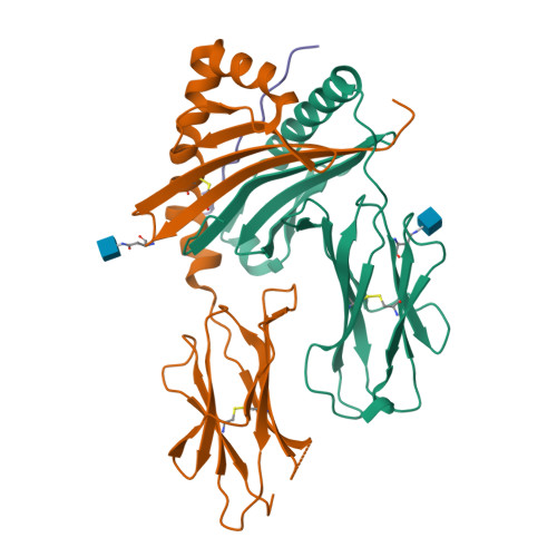
Crystal structure of MHC class II I-Ab in complex with a human CLIP peptide: Prediction of an I-Ab peptide-binding motif

Zhu, Y.Rudensky, A.Y.Teyton, A.L.Wilson, I.A.

(2003) J Mol Biol 326: 1157-1174

  • DOI: https://doi.org/10.1016/s0022-2836(02)01437-7
  • Primary Citation of Related Structures:  
    1MUJ

  • PubMed Abstract: 

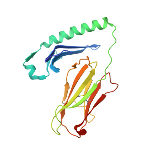
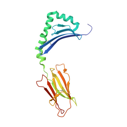
    Association between the class II major histocompatibility complex (MHC) and the class II invariant chain-associated peptide (CLIP) occurs naturally as an intermediate step in the MHC class II processing pathway. Here, we report the crystal structure of the murine class II MHC molecule I-A(b) in complex with human CLIP at 2.15A resolution. The structure of I-A(b) accounts, via the peptide-binding groove's unique physicochemistry, for the distinct peptide repertoire bound by this allele. CLIP adopts a similar conformation to peptides bound by other I-A alleles, reinforcing the notion that CLIP is presented as a conventional peptide antigen. When compared to the related HLA-DR3/CLIP complex structure, the CLIP peptide displays a slightly different conformation and distinct interaction pattern with residues in I-A(b). In addition, after examining the published sequences of peptides presented by I-A(b), we discuss the possibility of predicting peptide alignment in the I-A(b) binding groove using a simple scoring matrix.


  • Organizational Affiliation

    Department of Molecular Biology, The Scripps Research Institute, 10550 North Torrey Pines Road, La Jolla, CA 92037, USA.


Macromolecules
Find similar proteins by:  (by identity cutoff)  |  3D Structure
Entity ID: 1
MoleculeChains Sequence LengthOrganismDetailsImage
H-2 CLASS II HISTOCOMPATIBILITY ANTIGEN, A-B ALPHA CHAIN190Mus musculusMutation(s): 0 
UniProt
Find proteins for P14434 (Mus musculus)
Explore P14434 
Go to UniProtKB:  P14434
Entity Groups  
Sequence Clusters30% Identity50% Identity70% Identity90% Identity95% Identity100% Identity
UniProt GroupP14434
Glycosylation
Glycosylation Sites: 1
Sequence Annotations
Expand
  • Reference Sequence
Find similar proteins by:  (by identity cutoff)  |  3D Structure
Entity ID: 2
MoleculeChains Sequence LengthOrganismDetailsImage
H-2 CLASS II HISTOCOMPATIBILITY ANTIGEN, A BETA CHAIN197Mus musculusMutation(s): 0 
UniProt
Find proteins for P14483 (Mus musculus)
Explore P14483 
Go to UniProtKB:  P14483
Entity Groups  
Sequence Clusters30% Identity50% Identity70% Identity90% Identity95% Identity100% Identity
UniProt GroupP14483
Glycosylation
Glycosylation Sites: 1
Sequence Annotations
Expand
  • Reference Sequence
Find similar proteins by:  (by identity cutoff)  |  3D Structure
Entity ID: 3
MoleculeChains Sequence LengthOrganismDetailsImage
CLIP peptide36Homo sapiensMutation(s): 0 
UniProt & NIH Common Fund Data Resources
Find proteins for P04233 (Homo sapiens)
Explore P04233 
Go to UniProtKB:  P04233
PHAROS:  P04233
GTEx:  ENSG00000019582 
Entity Groups  
Sequence Clusters30% Identity50% Identity70% Identity90% Identity95% Identity100% Identity
UniProt GroupP04233
Sequence Annotations
Expand
  • Reference Sequence
Experimental Data & Validation

Experimental Data

  • Method: X-RAY DIFFRACTION
  • Resolution: 2.15 Å
  • R-Value Free:  0.246 (Depositor), 0.245 (DCC) 
  • R-Value Work:  0.220 (Depositor), 0.223 (DCC) 
  • R-Value Observed: 0.220 (Depositor) 
Space Group: C 1 2 1
Unit Cell:
Length ( Å )Angle ( ˚ )
a = 63.647α = 90
b = 88.065β = 92.48
c = 83.74γ = 90
Software Package:
Software NamePurpose
CNSrefinement
DENZOdata reduction
SCALEPACKdata scaling
MOLREPphasing

Structure Validation

View Full Validation Report


Ligand Structure Quality Assessment 

Created with Raphaël 2.3.0Worse 01 BetterLigand structure goodness of fit to experimental dataBest fitted NAGClick on this verticalbar to view details

Entry History 

Deposition Data

Revision History  (Full details and data files)

  • Version 1.0: 2003-02-18
    Type: Initial release
  • Version 1.1: 2008-04-28
    Changes: Version format compliance
  • Version 1.2: 2011-07-13
    Changes: Non-polymer description, Version format compliance
  • Version 1.3: 2020-07-29
    Type: Remediation
    Reason: Carbohydrate remediation
    Changes: Advisory, Data collection, Database references, Derived calculations, Structure summary
  • Version 1.4: 2024-10-30
    Changes: Advisory, Data collection, Database references, Structure summary