1N78

Crystal structure of Thermus thermophilus glutamyl-tRNA synthetase complexed with tRNA(Glu) and glutamol-AMP.


Experimental Data Snapshot

  • Method: X-RAY DIFFRACTION
  • Resolution: 2.10 Å
  • R-Value Free: 0.259 
  • R-Value Work: 0.219 
  • R-Value Observed: 0.220 

Starting Model: experimental
View more details

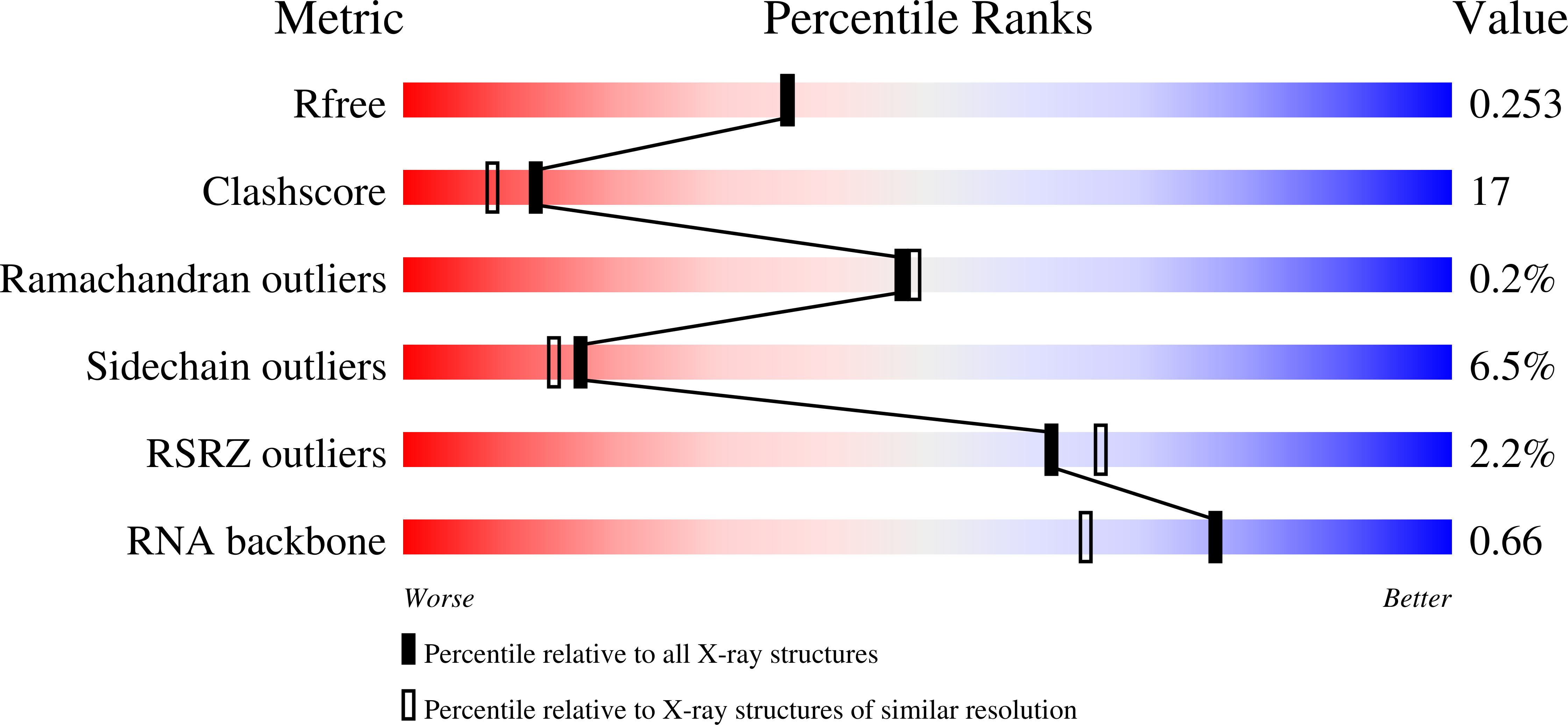
wwPDB Validation   3D Report Full Report


Ligand Structure Quality Assessment 


This is version 1.3 of the entry. See complete history


Literature

ATP binding by glutamyl-tRNA synthetase is switched to the productive mode by tRNA binding

Sekine, S.Nureki, O.Dubois, D.Y.Bernier, S.Chenevert, R.Lapointe, J.Vassylyev, D.G.Yokoyama, S.

(2003) EMBO J 22: 676-688

  • DOI: https://doi.org/10.1093/emboj/cdg053
  • Primary Citation of Related Structures:  
    1J09, 1N75, 1N77, 1N78

  • PubMed Abstract: 

    Aminoacyl-tRNA synthetases catalyze the formation of an aminoacyl-AMP from an amino acid and ATP, prior to the aminoacyl transfer to tRNA. A subset of aminoacyl-tRNA synthetases, including glutamyl-tRNA synthetase (GluRS), have a regulation mechanism to avoid aminoacyl-AMP formation in the absence of tRNA. In this study, we determined the crystal structure of the 'non-productive' complex of Thermus thermophilus GluRS, ATP and L-glutamate, together with those of the GluRS.ATP, GluRS.tRNA.ATP and GluRS.tRNA.GoA (a glutamyl-AMP analog) complexes. In the absence of tRNA(Glu), ATP is accommodated in a 'non-productive' subsite within the ATP-binding site, so that the ATP alpha-phosphate and the glutamate alpha-carboxyl groups in GluRS. ATP.Glu are too far from each other (6.2 A) to react. In contrast, the ATP-binding mode in GluRS.tRNA. ATP is dramatically different from those in GluRS.ATP.Glu and GluRS.ATP, but corresponds to the AMP moiety binding mode in GluRS.tRNA.GoA (the 'productive' subsite). Therefore, tRNA binding to GluRS switches the ATP-binding mode. The interactions of the three tRNA(Glu) regions with GluRS cause conformational changes around the ATP-binding site, and allow ATP to bind to the 'productive' subsite.


  • Organizational Affiliation

    Cellular Signaling Laboratory and Structurome Group, RIKEN Harima Institute at SPring-8, 1-1-1 Kouto, Mikazuki-cho, Sayo, Hyogo 679-5148, Japan.


Macromolecules

Find similar proteins by:  (by identity cutoff)  |  3D Structure
Entity ID: 2
MoleculeChains Sequence LengthOrganismDetailsImage
Glutamyl-tRNA synthetaseC [auth A],
D [auth B]
468Thermus thermophilusMutation(s): 0 
EC: 6.1.1.17
UniProt
Find proteins for P27000 (Thermus thermophilus (strain ATCC 27634 / DSM 579 / HB8))
Explore P27000 
Go to UniProtKB:  P27000
Entity Groups  
Sequence Clusters30% Identity50% Identity70% Identity90% Identity95% Identity100% Identity
UniProt GroupP27000
Sequence Annotations
Expand
  • Reference Sequence
Find similar nucleic acids by:  (by identity cutoff)  |  3D Structure
Entity ID: 1
MoleculeChains LengthOrganismImage
tRNA(Glu)A [auth C],
B [auth D]
75N/A
Sequence Annotations
Expand
  • Reference Sequence
Experimental Data & Validation

Experimental Data

  • Method: X-RAY DIFFRACTION
  • Resolution: 2.10 Å
  • R-Value Free: 0.259 
  • R-Value Work: 0.219 
  • R-Value Observed: 0.220 
  • Space Group: C 2 2 21
Unit Cell:
Length ( Å )Angle ( ˚ )
a = 110.492α = 90
b = 219.87β = 90
c = 135.119γ = 90
Software Package:
Software NamePurpose
HKL-2000data collection
SCALEPACKdata scaling
CNSrefinement
HKL-2000data reduction
CNSphasing

Structure Validation

View Full Validation Report


Ligand Structure Quality Assessment 


Entry History 

Revision History  (Full details and data files)

  • Version 1.0: 2003-02-25
    Type: Initial release
  • Version 1.1: 2008-04-28
    Changes: Version format compliance
  • Version 1.2: 2011-07-13
    Changes: Version format compliance
  • Version 1.3: 2023-10-25
    Changes: Data collection, Database references, Derived calculations, Refinement description