3R6N

Crystal structure of a rigid four spectrin repeat fragment of the human desmoplakin plakin domain


Experimental Data Snapshot

  • Method: X-RAY DIFFRACTION
  • Resolution: 2.95 Å
  • R-Value Free: 
    0.265 (Depositor), 0.260 (DCC) 
  • R-Value Work: 
    0.216 (Depositor), 0.210 (DCC) 
  • R-Value Observed: 
    0.221 (Depositor) 

wwPDB Validation   3D Report Full Report


Ligand Structure Quality Assessment 

Created with Raphaël 2.3.0Worse 01 BetterLigand structure goodness of fit to experimental dataBest fitted D1DClick on this verticalbar to view details

This is version 1.2 of the entry. See complete history


Literature

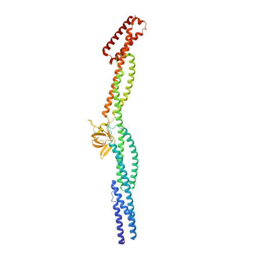
Crystal structure of a rigid four-spectrin-repeat fragment of the human desmoplakin plakin domain.

Choi, H.J.Weis, W.I.

(2011) J Mol Biol 409: 800-812

  • DOI: https://doi.org/10.1016/j.jmb.2011.04.046
  • Primary Citation of Related Structures:  
    3R6N

  • PubMed Abstract: 

    The plakin protein family serves to connect cell-cell and cell-matrix adhesion molecules to the intermediate filament cytoskeleton. Desmoplakin (DP) is an integral part of desmosomes, where it links desmosomal cadherins to the intermediate filaments. The 1056-amino-acid N-terminal region of DP contains a plakin domain common to members of the plakin family. Plakin domains contain multiple copies of spectrin repeats (SRs). We determined the crystal structure of a fragment of DP, residues 175-630, consisting of four SRs and an inserted SH3 domain. The four repeats form an elongated, rigid structure. The SH3 domain is present in a loop between two helices of an SR and interacts extensively with the preceding SR in a manner that appears to limit inter-repeat flexibility. The intimate intramolecular association of the SH3 domain with the preceding SR is also observed in plectin, another plakin protein, but not in α-spectrin, suggesting that the SH3 domain of plakins contributes to the stability and rigidity of this subfamily of SR-containing proteins.


  • Organizational Affiliation

    Department of Structural Biology, Stanford University School of Medicine, Stanford, CA 94305, USA.


Macromolecules
Find similar proteins by:  (by identity cutoff)  |  3D Structure
Entity ID: 1
MoleculeChains Sequence LengthOrganismDetailsImage
Desmoplakin
A, B
450Homo sapiensMutation(s): 0 
Gene Names: DSP
UniProt & NIH Common Fund Data Resources
Find proteins for P15924 (Homo sapiens)
Explore P15924 
Go to UniProtKB:  P15924
PHAROS:  P15924
GTEx:  ENSG00000096696 
Entity Groups  
Sequence Clusters30% Identity50% Identity70% Identity90% Identity95% Identity100% Identity
UniProt GroupP15924
Sequence Annotations
Expand
  • Reference Sequence
Experimental Data & Validation

Experimental Data

  • Method: X-RAY DIFFRACTION
  • Resolution: 2.95 Å
  • R-Value Free:  0.265 (Depositor), 0.260 (DCC) 
  • R-Value Work:  0.216 (Depositor), 0.210 (DCC) 
  • R-Value Observed: 0.221 (Depositor) 
Space Group: C 1 2 1
Unit Cell:
Length ( Å )Angle ( ˚ )
a = 157.52α = 90
b = 82.52β = 125.54
c = 139.98γ = 90
Software Package:
Software NamePurpose
MOSFLMdata reduction
SCALAdata scaling
PHASERphasing
SnBphasing
RESOLVEphasing
PHENIXrefinement
PDB_EXTRACTdata extraction

Structure Validation

View Full Validation Report


Ligand Structure Quality Assessment 

Created with Raphaël 2.3.0Worse 01 BetterLigand structure goodness of fit to experimental dataBest fitted D1DClick on this verticalbar to view details

Entry History 

Deposition Data

Revision History  (Full details and data files)

  • Version 1.0: 2011-05-11
    Type: Initial release
  • Version 1.1: 2011-07-13
    Changes: Version format compliance
  • Version 1.2: 2024-02-21
    Changes: Data collection, Database references, Derived calculations