3RSN

Crystal Structure of the N-terminal region of Human Ash2L


Experimental Data Snapshot

  • Method: X-RAY DIFFRACTION
  • Resolution: 2.10 Å
  • R-Value Free: 0.254 
  • R-Value Work: 0.213 
  • R-Value Observed: 0.217 

wwPDB Validation   3D Report Full Report


This is version 1.2 of the entry. See complete history


Literature

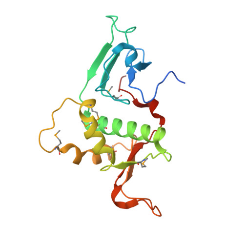
Crystal structure of the N-terminal region of human Ash2L shows a winged-helix motif involved in DNA binding.

Chen, Y.Wan, B.Wang, K.C.Cao, F.Yang, Y.Protacio, A.Dou, Y.Chang, H.Y.Lei, M.

(2011) EMBO Rep 12: 797-803

  • DOI: https://doi.org/10.1038/embor.2011.101
  • Primary Citation of Related Structures:  
    3RSN

  • PubMed Abstract: 

    Ash2L is a core component of the MLL family histone methyltransferases and has an important role in regulating the methylation of histone H3 on lysine 4. Here, we report the crystal structure of the N-terminal domain of Ash2L and reveal a new function of Ash2L. The structure shows that Ash2L contains an atypical PHD finger that does not have histone tail-binding activity. Unexpectedly, the structure shows a previously unrecognized winged-helix motif that directly binds to DNA. The DNA-binding-deficient mutants of Ash2L reduced Ash2L localization to the HOX locus. Strikingly, a single mutation in Ash2L(WH) (K131A) breaks the chromatin domain boundary, suggesting that Ash2L also has a role in chromosome demarcation.


  • Organizational Affiliation

    Howard Hughes Medical Institute, 1150 West, Medical Center Drive, Ann Arbor, Michigan 48109, USA.


Macromolecules
Find similar proteins by:  (by identity cutoff)  |  3D Structure
Entity ID: 1
MoleculeChains Sequence LengthOrganismDetailsImage
Set1/Ash2 histone methyltransferase complex subunit ASH2177Homo sapiensMutation(s): 0 
Gene Names: ASH2LASH2L1
UniProt & NIH Common Fund Data Resources
Find proteins for Q9UBL3 (Homo sapiens)
Explore Q9UBL3 
Go to UniProtKB:  Q9UBL3
PHAROS:  Q9UBL3
GTEx:  ENSG00000129691 
Entity Groups  
Sequence Clusters30% Identity50% Identity70% Identity90% Identity95% Identity100% Identity
UniProt GroupQ9UBL3
Sequence Annotations
Expand
  • Reference Sequence
Small Molecules
Ligands 1 Unique
IDChains Name / Formula / InChI Key2D Diagram3D Interactions
ZN
Query on ZN

Download Ideal Coordinates CCD File 
B [auth A]ZINC ION
Zn
PTFCDOFLOPIGGS-UHFFFAOYSA-N
Modified Residues  1 Unique
IDChains TypeFormula2D DiagramParent
MSE
Query on MSE
A
L-PEPTIDE LINKINGC5 H11 N O2 SeMET
Experimental Data & Validation

Experimental Data

  • Method: X-RAY DIFFRACTION
  • Resolution: 2.10 Å
  • R-Value Free: 0.254 
  • R-Value Work: 0.213 
  • R-Value Observed: 0.217 
  • Space Group: P 31 2 1
Unit Cell:
Length ( Å )Angle ( ˚ )
a = 49.984α = 90
b = 49.984β = 90
c = 165.519γ = 120
Software Package:
Software NamePurpose
MD2data collection
SHARPphasing
PHENIXrefinement
HKL-2000data reduction
HKL-2000data scaling

Structure Validation

View Full Validation Report


Entry History 

Deposition Data

Revision History  (Full details and data files)

  • Version 1.0: 2011-06-22
    Type: Initial release
  • Version 1.1: 2011-07-13
    Changes: Version format compliance
  • Version 1.2: 2011-12-14
    Changes: Database references