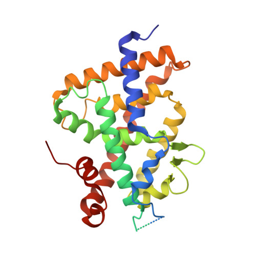
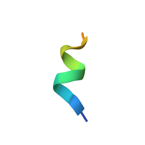
4RUJ

Crystal structure of zVDR L337H mutant-VD complex


Experimental Data Snapshot

  • Method: X-RAY DIFFRACTION
  • Resolution: 2.35 Å
  • R-Value Free: 0.255 
  • R-Value Work: 0.200 
  • R-Value Observed: 0.203 

wwPDB Validation   3D Report Full Report


Ligand Structure Quality Assessment 


This is version 1.2 of the entry. See complete history


Literature

A vitamin D receptor selectively activated by gemini analogs reveals ligand dependent and independent effects.

Huet, T.Laverny, G.Ciesielski, F.Molnar, F.Ramamoorthy, T.G.Belorusova, A.Y.Antony, P.Potier, N.Metzger, D.Moras, D.Rochel, N.

(2015) Cell Rep 10: 516-526

  • DOI: https://doi.org/10.1016/j.celrep.2014.12.045
  • Primary Citation of Related Structures:  
    4RUJ, 4RUO, 4RUP

  • PubMed Abstract: 

    The bioactive form of vitamin D [1,25(OH)2D3] regulates mineral and bone homeostasis and exerts potent anti-inflammatory and antiproliferative properties through binding to the vitamin D receptor (VDR). The 3D structures of the VDR ligand-binding domain with 1,25(OH)2D3 or gemini analogs unveiled the molecular mechanism underlying ligand recognition. On the basis of structure-function correlations, we generated a point-mutated VDR (VDR(gem)) that is unresponsive to 1,25(OH)2D3, but the activity of which is efficiently induced by the gemini ligands. Moreover, we show that many VDR target genes are repressed by unliganded VDR(gem) and that mineral ion and bone homeostasis are more impaired in VDR(gem) mice than in VDR null mice, demonstrating that mutations abolishing VDR ligand binding result in more severe skeletal defects than VDR null mutations. As gemini ligands induce VDR(gem) transcriptional activity in mice and normalize their serum calcium levels, VDR(gem) is a powerful tool to further unravel both liganded and unliganded VDR signaling.


  • Organizational Affiliation

    Department of Integrative Structural Biology, Institut de Génétique et de Biologie Moléculaire et Cellulaire (IGBMC), Institut National de la Santé et de la Recherche Médicale (INSERM) U964, Centre National de la Recherche Scientifique (CNRS) UMR 7104, Université de Strasbourg, 67404 Illkirch, France.


Macromolecules
Find similar proteins by:  (by identity cutoff)  |  3D Structure
Entity ID: 1
MoleculeChains Sequence LengthOrganismDetailsImage
Vitamin D3 receptor A302Danio rerioMutation(s): 1 
Gene Names: vdranr1i1avdr
UniProt
Find proteins for Q9PTN2 (Danio rerio)
Explore Q9PTN2 
Go to UniProtKB:  Q9PTN2
Entity Groups  
Sequence Clusters30% Identity50% Identity70% Identity90% Identity95% Identity100% Identity
UniProt GroupQ9PTN2
Sequence Annotations
Expand
  • Reference Sequence

Find similar proteins by:  Sequence   |   3D Structure  

Entity ID: 2
MoleculeChains Sequence LengthOrganismDetailsImage
Nuclear receptor coactivator 115Homo sapiensMutation(s): 0 
EC: 2.3.1.48
UniProt & NIH Common Fund Data Resources
Find proteins for Q15788 (Homo sapiens)
Explore Q15788 
Go to UniProtKB:  Q15788
PHAROS:  Q15788
GTEx:  ENSG00000084676 
Entity Groups  
Sequence Clusters30% Identity50% Identity70% Identity90% Identity95% Identity100% Identity
UniProt GroupQ15788
Sequence Annotations
Expand
  • Reference Sequence
Small Molecules
Ligands 1 Unique
IDChains Name / Formula / InChI Key2D Diagram3D Interactions
VDX
Query on VDX

Download Ideal Coordinates CCD File 
C [auth A]5-{2-[1-(5-HYDROXY-1,5-DIMETHYL-HEXYL)-7A-METHYL-OCTAHYDRO-INDEN-4-YLIDENE]-ETHYLIDENE}-4-METHYLENE-CYCLOHEXANE-1,3-DIOL
C27 H44 O3
GMRQFYUYWCNGIN-NKMMMXOESA-N
Binding Affinity Annotations 
IDSourceBinding Affinity
VDX BindingDB:  4RUJ Kd: 1200 (nM) from 1 assay(s)
EC50: 5.5 (nM) from 1 assay(s)
Experimental Data & Validation

Experimental Data

  • Method: X-RAY DIFFRACTION
  • Resolution: 2.35 Å
  • R-Value Free: 0.255 
  • R-Value Work: 0.200 
  • R-Value Observed: 0.203 
  • Space Group: P 65 2 2
Unit Cell:
Length ( Å )Angle ( ˚ )
a = 65.851α = 90
b = 65.851β = 90
c = 264.373γ = 120
Software Package:
Software NamePurpose
PHENIXrefinement
CNSrefinement
ADSCdata collection
HKL-2000data reduction
HKL-2000data scaling
CNSphasing

Structure Validation

View Full Validation Report


Ligand Structure Quality Assessment 


Entry History 

Deposition Data

Revision History  (Full details and data files)

  • Version 1.0: 2015-10-07
    Type: Initial release
  • Version 1.1: 2019-07-17
    Changes: Advisory, Data collection, Refinement description
  • Version 1.2: 2024-02-28
    Changes: Advisory, Data collection, Database references, Derived calculations