5C1M

Crystal structure of active mu-opioid receptor bound to the agonist BU72


Experimental Data Snapshot

  • Method: X-RAY DIFFRACTION
  • Resolution: 2.07 Å
  • R-Value Free: 
    0.229 (Depositor), 0.230 (DCC) 
  • R-Value Work: 
    0.199 (Depositor), 0.200 (DCC) 
  • R-Value Observed: 
    0.200 (Depositor) 

Starting Models: experimental
View more details

wwPDB Validation   3D Report Full Report


Ligand Structure Quality Assessment 

Created with Raphaël 2.3.0Worse 01 BetterLigand structure goodness of fit to experimental dataBest fitted VF1Click on this verticalbar to view details

This is version 2.1 of the entry. See complete history

Re-refinement Note

A newer entry is available that reflects an alternative modeling of the original data: 8E0G


Literature

Structural insights into mu-opioid receptor activation.

Huang, W.Manglik, A.Venkatakrishnan, A.J.Laeremans, T.Feinberg, E.N.Sanborn, A.L.Kato, H.E.Livingston, K.E.Thorsen, T.S.Kling, R.C.Granier, S.Gmeiner, P.Husbands, S.M.Traynor, J.R.Weis, W.I.Steyaert, J.Dror, R.O.Kobilka, B.K.

(2015) Nature 524: 315-321

  • DOI: https://doi.org/10.1038/nature14886
  • Primary Citation of Related Structures:  
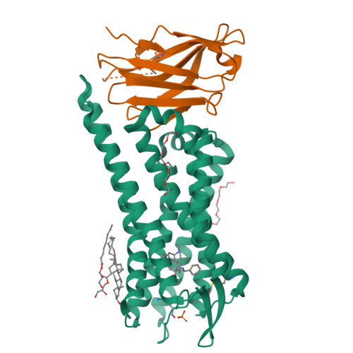
    5C1M

  • PubMed Abstract: 

    Activation of the μ-opioid receptor (μOR) is responsible for the efficacy of the most effective analgesics. To shed light on the structural basis for μOR activation, here we report a 2.1 Å X-ray crystal structure of the murine μOR bound to the morphinan agonist BU72 and a G protein mimetic camelid antibody fragment. The BU72-stabilized changes in the μOR binding pocket are subtle and differ from those observed for agonist-bound structures of the β2-adrenergic receptor (β2AR) and the M2 muscarinic receptor. Comparison with active β2AR reveals a common rearrangement in the packing of three conserved amino acids in the core of the μOR, and molecular dynamics simulations illustrate how the ligand-binding pocket is conformationally linked to this conserved triad. Additionally, an extensive polar network between the ligand-binding pocket and the cytoplasmic domains appears to play a similar role in signal propagation for all three G-protein-coupled receptors.


  • Organizational Affiliation

    Department of Molecular and Cellular Physiology, Stanford University School of Medicine, 279 Campus Drive, Stanford, California 94305, USA.


Macromolecules
Find similar proteins by:  (by identity cutoff)  |  3D Structure
Entity ID: 1
MoleculeChains Sequence LengthOrganismDetailsImage
Mu-type opioid receptor296Mus musculusMutation(s): 0 
Gene Names: Oprm1MorOprm
Membrane Entity: Yes 
UniProt
Find proteins for P42866 (Mus musculus)
Explore P42866 
Go to UniProtKB:  P42866
Entity Groups  
Sequence Clusters30% Identity50% Identity70% Identity90% Identity95% Identity100% Identity
UniProt GroupP42866
Sequence Annotations
Expand
  • Reference Sequence
Find similar proteins by:  (by identity cutoff)  |  3D Structure
Entity ID: 2
MoleculeChains Sequence LengthOrganismDetailsImage
Nanobody 39125Lama glamaMutation(s): 0 
Entity Groups  
Sequence Clusters30% Identity50% Identity70% Identity90% Identity95% Identity100% Identity
Sequence Annotations
Expand
  • Reference Sequence
Small Molecules
Ligands 5 Unique
IDChains Name / Formula / InChI Key2D Diagram3D Interactions
VF1 (Subject of Investigation/LOI)
Query on VF1

Download Ideal Coordinates CCD File 
I [auth A](2R,3S,3aR,5aR,6R,11bR,11cS)-3a-methoxy-3,14-dimethyl-2-phenyl-2,3,3a,6,7,11c-hexahydro-1H-6,11b-(epiminoethano)-3,5a-methanonaphtho[2,1-g]indol-10-ol
C28 H32 N2 O2
RGJHUVJQGAAZLK-GKTFKBBASA-N
CLR
Query on CLR

Download Ideal Coordinates CCD File 
E [auth A]CHOLESTEROL
C27 H46 O
HVYWMOMLDIMFJA-DPAQBDIFSA-N
OLC
Query on OLC

Download Ideal Coordinates CCD File 
C [auth A],
D [auth A]
(2R)-2,3-dihydroxypropyl (9Z)-octadec-9-enoate
C21 H40 O4
RZRNAYUHWVFMIP-GDCKJWNLSA-N
P6G
Query on P6G

Download Ideal Coordinates CCD File 
G [auth A],
H [auth A]
HEXAETHYLENE GLYCOL
C12 H26 O7
IIRDTKBZINWQAW-UHFFFAOYSA-N
PO4
Query on PO4

Download Ideal Coordinates CCD File 
F [auth A]PHOSPHATE ION
O4 P
NBIIXXVUZAFLBC-UHFFFAOYSA-K
Modified Residues  1 Unique
IDChains TypeFormula2D DiagramParent
YCM
Query on YCM
A
L-PEPTIDE LINKINGC5 H10 N2 O3 SCYS
Experimental Data & Validation

Experimental Data

  • Method: X-RAY DIFFRACTION
  • Resolution: 2.07 Å
  • R-Value Free:  0.229 (Depositor), 0.230 (DCC) 
  • R-Value Work:  0.199 (Depositor), 0.200 (DCC) 
  • R-Value Observed: 0.200 (Depositor) 
Space Group: I 21 21 21
Unit Cell:
Length ( Å )Angle ( ˚ )
a = 44.43α = 90
b = 144β = 90
c = 209.9γ = 90
Software Package:
Software NamePurpose
PHENIXrefinement
PHENIXrefinement
XSCALEdata scaling
PDB_EXTRACTdata extraction
PHASERphasing
XDSdata reduction

Structure Validation

View Full Validation Report


Ligand Structure Quality Assessment 

Created with Raphaël 2.3.0Worse 01 BetterLigand structure goodness of fit to experimental dataBest fitted VF1Click on this verticalbar to view details

Entry History & Funding Information

Deposition Data


Funding OrganizationLocationGrant Number
National Institutes of Health/National Institute on Drug Abuse (NIH/NIDA)United StatesR37DA036246
National Institutes of Health/National Institute of General Medical Sciences (NIH/NIGMS)United StatesR01GM083118

Revision History  (Full details and data files)

  • Version 1.0: 2015-08-05
    Type: Initial release
  • Version 1.1: 2015-08-19
    Changes: Database references
  • Version 1.2: 2015-08-26
    Changes: Data collection
  • Version 1.3: 2015-09-02
    Changes: Database references
  • Version 1.4: 2017-09-06
    Changes: Author supporting evidence, Database references, Derived calculations
  • Version 1.5: 2019-12-11
    Changes: Author supporting evidence
  • Version 2.0: 2021-02-10
    Type: Coordinate replacement
    Reason: Chirality error
    Changes: Advisory, Atomic model, Author supporting evidence, Data collection, Derived calculations, Non-polymer description, Other, Refinement description, Structure summary
  • Version 2.1: 2023-09-27
    Changes: Data collection, Database references, Refinement description