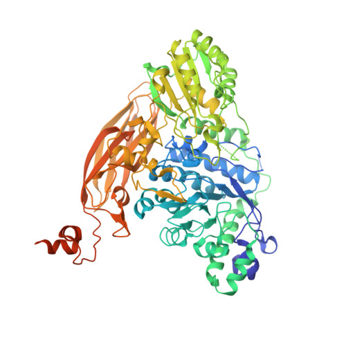
Structural and biochemical characterization of a GH3 beta-glucosidase from the probiotic bacteria Bifidobacterium adolescentis.
Florindo, R.N., Souza, V.P., Manzine, L.R., Camilo, C.M., Marana, S.R., Polikarpov, I., Nascimento, A.S.(2018) Biochimie 148: 107-115
- PubMed: 29555372
- DOI: https://doi.org/10.1016/j.biochi.2018.03.007
- Primary Citation of Related Structures:
5WAB - PubMed Abstract:
Bifidobacterium is an important genus of probiotic bacteria colonizing the human gut. These bacteria can uptake oligosaccharides for the fermentative metabolism of hexoses and pentoses, producing lactate, acetate as well as short-chain fatty acids and propionate. These end-products are known to have important effects on human health. β-glucosidases (EC 3.2.1.21) are pivotal enzymes for the metabolism and homeostasis of Bifidobacterium, since they hydrolyze small and soluble saccharides, typically producing glucose. Here we describe the cloning, expression, biochemical characterization and the first X-ray structure of a GH3 β-glucosidase from the probiotic bacteria Bifidobacterium adolescentis (BaBgl3). The purified BaBgl3 showed a maximal activity at 45 °C and pH 6.5. Under the optimum conditions, BaBgl3 is highly active on 4-nitrophenyl-β-d-glucopyranoside (pNPG) and, at a lesser degree, on 4-nitrophenyl-β-d-xylopyranoside (pNPX, about 32% of the activity observed for pNPG). The 2.4 Å resolution crystal structure of BaBgl3 revealed a three-domain structure composed of a TIM barrel domain, which together with α/β sandwich domain accommodate the active site and a third C-terminal fibronectin type III (FnIII) domain with unknown function. Modeling of the substrate in the active site indicates that an aspartate interacts with the hydroxyl group of the C6 present in pNPG but absent in pNPX, which explains the substrate preference. Finally, the enzyme is significantly stabilized by glycerol and galactose, resulting in considerable increase in the enzyme activity and its lifetime. The structural and biochemical studies presented here provide a deeper understanding of the molecular mechanisms of complex carbohydrates degradation utilized by probiotic bacteria as well as for the development of new prebiotic oligosaccharides.
Organizational Affiliation:
Instituto de Física de São Carlos, Universidade de São Paulo, Av. Trabalhador Saocarlense, 400. Pq. Arnold Schmidt, São Carlos, SP 13566-590, Brazil.


