7A5D

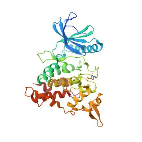
Structure of DYRK1A in complex with compound 16


Experimental Data Snapshot

  • Method: X-RAY DIFFRACTION
  • Resolution: 1.80 Å
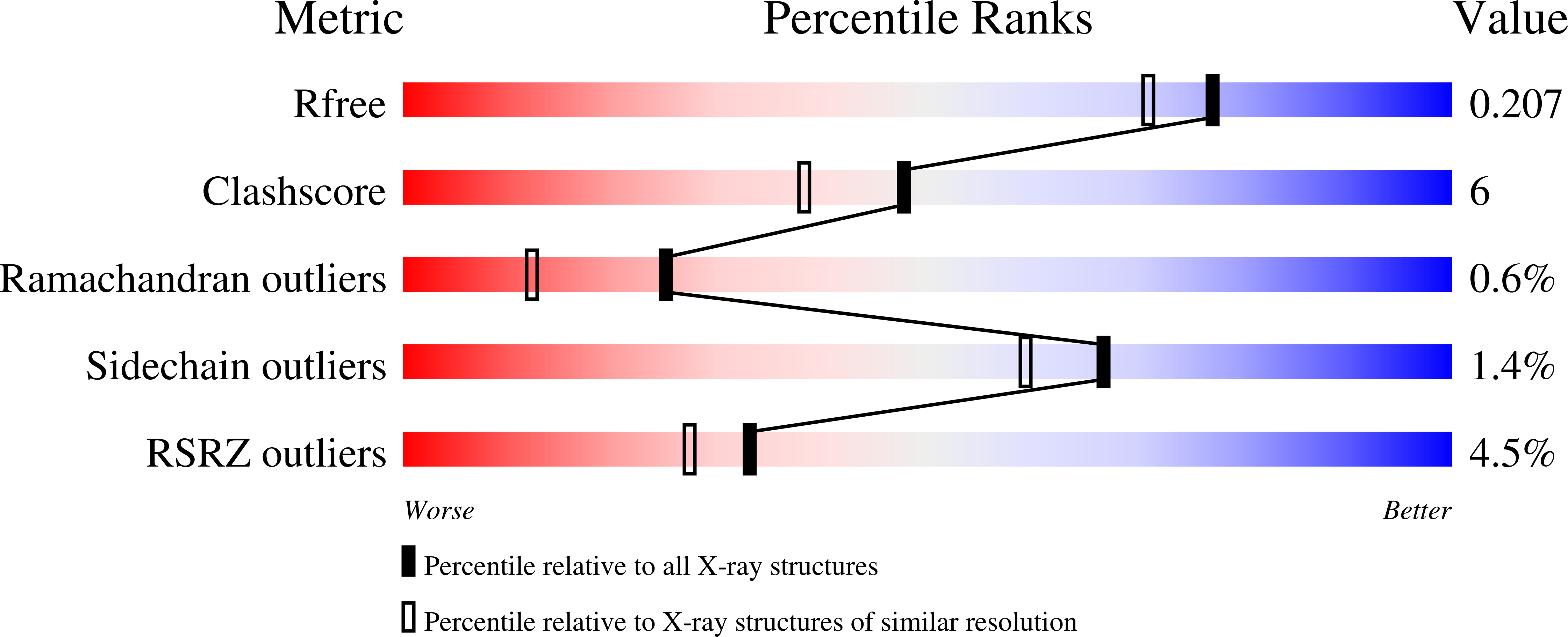
  • R-Value Free: 0.198 
  • R-Value Work: 0.167 
  • R-Value Observed: 0.169 

Starting Model: experimental
View more details

wwPDB Validation   3D Report Full Report


Ligand Structure Quality Assessment 


This is version 1.3 of the entry. See complete history


Literature

Fragment-Derived Selective Inhibitors of Dual-Specificity Kinases DYRK1A and DYRK1B.

Lee Walmsley, D.Murray, J.B.Dokurno, P.Massey, A.J.Benwell, K.Fiumana, A.Foloppe, N.Ray, S.Smith, J.Surgenor, A.E.Edmonds, T.Demarles, D.Burbridge, M.Cruzalegui, F.Kotschy, A.Hubbard, R.E.

(2021) J Med Chem 64: 8971-8991

  • DOI: https://doi.org/10.1021/acs.jmedchem.1c00024
  • Primary Citation of Related Structures:  
    7A4O, 7A4R, 7A4S, 7A4W, 7A4Z, 7A51, 7A52, 7A53, 7A55, 7A5B, 7A5D, 7A5L, 7A5N

  • PubMed Abstract: 

    The serine/threonine kinase DYRK1A has been implicated in regulation of a variety of cellular processes associated with cancer progression, including cell cycle control, DNA damage repair, protection from apoptosis, cell differentiation, and metastasis. In addition, elevated-level DYRK1A activity has been associated with increased severity of symptoms in Down's syndrome. A selective inhibitor of DYRK1A could therefore be of therapeutic benefit. We have used fragment and structure-based discovery methods to identify a highly selective, well-tolerated, brain-penetrant DYRK1A inhibitor which showed in vivo activity in a tumor model. The inhibitor provides a useful tool compound for further exploration of the effect of DYRK1A inhibition in models of disease.


  • Organizational Affiliation

    Vernalis (R&D) Ltd., Granta Park, Cambridge CB21 6GB, U.K.


Macromolecules
Find similar proteins by:  (by identity cutoff)  |  3D Structure
Entity ID: 1
MoleculeChains Sequence LengthOrganismDetailsImage
Dual specificity tyrosine-phosphorylation-regulated kinase 1A359Homo sapiensMutation(s): 0 
Gene Names: DYRK1ADYRKMNBMNBH
EC: 2.7.12.1 (PDB Primary Data), 2.7.11.23 (UniProt)
UniProt & NIH Common Fund Data Resources
Find proteins for Q13627 (Homo sapiens)
Explore Q13627 
Go to UniProtKB:  Q13627
PHAROS:  Q13627
GTEx:  ENSG00000157540 
Entity Groups  
Sequence Clusters30% Identity50% Identity70% Identity90% Identity95% Identity100% Identity
UniProt GroupQ13627
Sequence Annotations
Expand
  • Reference Sequence
Small Molecules
Modified Residues  2 Unique
IDChains TypeFormula2D DiagramParent
PTR
Query on PTR
A
L-PEPTIDE LINKINGC9 H12 N O6 PTYR
SEP
Query on SEP
A
L-PEPTIDE LINKINGC3 H8 N O6 PSER
Experimental Data & Validation

Experimental Data

  • Method: X-RAY DIFFRACTION
  • Resolution: 1.80 Å
  • R-Value Free: 0.198 
  • R-Value Work: 0.167 
  • R-Value Observed: 0.169 
  • Space Group: P 43 21 2
Unit Cell:
Length ( Å )Angle ( ˚ )
a = 81.696α = 90
b = 81.696β = 90
c = 123.601γ = 90
Software Package:
Software NamePurpose
REFMACrefinement
XDSdata reduction
XSCALEdata scaling
MOLREPphasing
PDB_EXTRACTdata extraction

Structure Validation

View Full Validation Report


Ligand Structure Quality Assessment 


Entry History 

Deposition Data

Revision History  (Full details and data files)

  • Version 1.0: 2021-06-30
    Type: Initial release
  • Version 1.1: 2021-07-21
    Changes: Database references
  • Version 1.2: 2024-01-31
    Changes: Data collection, Database references, Refinement description
  • Version 1.3: 2024-11-13
    Changes: Structure summary