7DHA

crystal structure of CD38 in complex with daratumumab


Experimental Data Snapshot

  • Method: X-RAY DIFFRACTION
  • Resolution: 2.55 Å
  • R-Value Free: 0.261 
  • R-Value Work: 0.207 
  • R-Value Observed: 0.211 

wwPDB Validation   3D Report Full Report


This is version 1.1 of the entry. See complete history


Literature

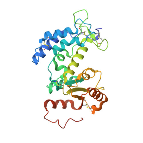
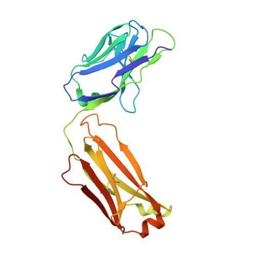
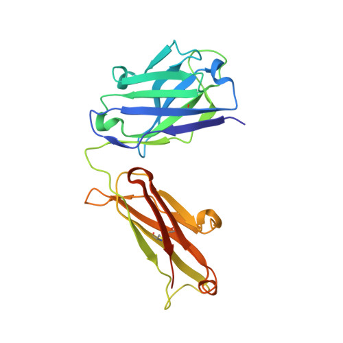
Crystal structure of CD38 in complex with daratumumab, a first-in-class anti-CD38 antibody drug for treating multiple myeloma.

Lee, H.T.Kim, Y.Park, U.B.Jeong, T.J.Lee, S.H.Heo, Y.S.

(2021) Biochem Biophys Res Commun 536: 26-31

  • DOI: https://doi.org/10.1016/j.bbrc.2020.12.048
  • Primary Citation of Related Structures:  
    7DHA

  • PubMed Abstract: 

    Multiple myeloma is a blood cancer characterized by the plasma cell malignancy in the bone marrow, resulting in the destruction of bone tissue. Recently, the US FDA approved two antibody drugs for the treatment of multiple myeloma, daratumumab and isatuximab, targeting CD38, a type II transmembrane glycoprotein highly expressed in plasma cells and multiple myeloma cells. Here, we report the crystal structure of CD38 in complex with the Fab fragment of daratumumab, providing its exact epitope on CD38 and the structural insights into the mechanism of action of the antibody drug. Daratumumab binds to a specific discontinuous region on CD38 that includes residues located opposite to the active site of CD38. All the six complementarity determining regions of daratumumab are involved in the CD38 interaction. The epitopes of daratumumab and isatuximab do not overlap at all and their bindings to CD38 induce different structural changes within the CD38 protein. This structural study can facilitate the design of improved biologics or effective combination therapies for the treatment of multiple myeloma.


  • Organizational Affiliation

    Department of Chemistry, Konkuk University, 120 Neungdong-ro, Gwangjin-gu, Seoul, 05029, Republic of Korea.


Macromolecules
Find similar proteins by:  (by identity cutoff)  |  3D Structure
Entity ID: 1
MoleculeChains Sequence LengthOrganismDetailsImage
ADP-ribosyl cyclase/cyclic ADP-ribose hydrolase 1265Homo sapiensMutation(s): 4 
Gene Names: CD38
EC: 3.2.2.6 (PDB Primary Data), 2.4.99.20 (PDB Primary Data)
UniProt & NIH Common Fund Data Resources
Find proteins for P28907 (Homo sapiens)
Explore P28907 
Go to UniProtKB:  P28907
PHAROS:  P28907
GTEx:  ENSG00000004468 
Entity Groups  
Sequence Clusters30% Identity50% Identity70% Identity90% Identity95% Identity100% Identity
UniProt GroupP28907
Sequence Annotations
Expand
  • Reference Sequence
Find similar proteins by:  (by identity cutoff)  |  3D Structure
Entity ID: 2
MoleculeChains Sequence LengthOrganismDetailsImage
Light chain214Homo sapiensMutation(s): 0 
Entity Groups  
Sequence Clusters30% Identity50% Identity70% Identity90% Identity95% Identity100% Identity
Sequence Annotations
Expand
  • Reference Sequence
Find similar proteins by:  (by identity cutoff)  |  3D Structure
Entity ID: 3
MoleculeChains Sequence LengthOrganismDetailsImage
Heavy Chain234Homo sapiensMutation(s): 0 
Entity Groups  
Sequence Clusters30% Identity50% Identity70% Identity90% Identity95% Identity100% Identity
Sequence Annotations
Expand
  • Reference Sequence
Experimental Data & Validation

Experimental Data

  • Method: X-RAY DIFFRACTION
  • Resolution: 2.55 Å
  • R-Value Free: 0.261 
  • R-Value Work: 0.207 
  • R-Value Observed: 0.211 
  • Space Group: P 1 2 1
Unit Cell:
Length ( Å )Angle ( ˚ )
a = 80.24α = 90
b = 54.16β = 98.878
c = 92.69γ = 90
Software Package:
Software NamePurpose
MxDCdata collection
PHENIXrefinement
XDSdata reduction
XDSdata scaling
MOLREPphasing

Structure Validation

View Full Validation Report


Entry History 

Deposition Data

Revision History  (Full details and data files)

  • Version 1.0: 2021-09-22
    Type: Initial release
  • Version 1.1: 2023-11-29
    Changes: Data collection, Refinement description