7DVF

Crystal structure of the computationally designed reDPBB_sym2 protein


Experimental Data Snapshot

  • Method: X-RAY DIFFRACTION
  • Resolution: 1.21 Å
  • R-Value Free: 0.195 
  • R-Value Work: 0.154 
  • R-Value Observed: 0.157 

wwPDB Validation   3D Report Full Report


This is version 1.2 of the entry. See complete history


Literature

Seven Amino Acid Types Suffice to Create the Core Fold of RNA Polymerase.

Yagi, S.Padhi, A.K.Vucinic, J.Barbe, S.Schiex, T.Nakagawa, R.Simoncini, D.Zhang, K.Y.J.Tagami, S.

(2021) J Am Chem Soc 143: 15998-16006

  • DOI: https://doi.org/10.1021/jacs.1c05367
  • Primary Citation of Related Structures:  
    7DBO, 7DG7, 7DG9, 7DI0, 7DI1, 7DU6, 7DU7, 7DVC, 7DVF, 7DVH, 7DWW, 7DXR, 7DXS, 7DXT, 7DXU, 7DXV, 7DXW, 7DXX, 7DXY, 7DXZ, 7DYC

  • PubMed Abstract: 

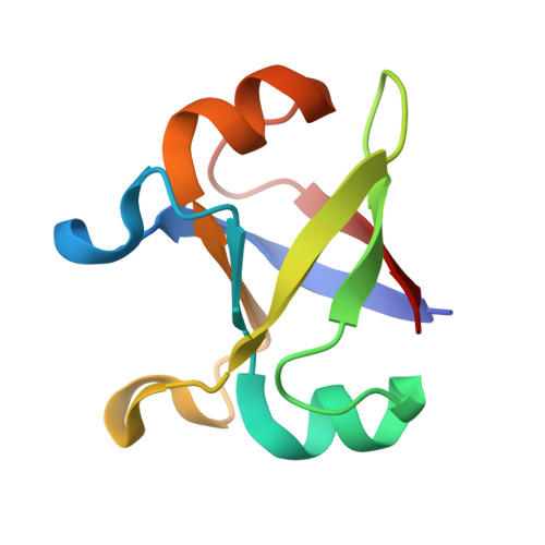
    The extant complex proteins must have evolved from ancient short and simple ancestors. The double-ψ β-barrel (DPBB) is one of the oldest protein folds and conserved in various fundamental enzymes, such as the core domain of RNA polymerase. Here, by reverse engineering a modern DPBB domain, we reconstructed its plausible evolutionary pathway started by "interlacing homodimerization" of a half-size peptide, followed by gene duplication and fusion. Furthermore, by simplifying the amino acid repertoire of the peptide, we successfully created the DPBB fold with only seven amino acid types (Ala, Asp, Glu, Gly, Lys, Arg, and Val), which can be coded by only GNN and ARR (R = A or G) codons in the modern translation system. Thus, the DPBB fold could have been materialized by the early translation system and genetic code.


  • Organizational Affiliation

    RIKEN Center for Biosystems Dynamics Research, 1-7-22 Suehiro-cho, Tsurumi-ku, Yokohama, Kanagawa 230-0045, Japan.


Macromolecules
Find similar proteins by:  (by identity cutoff)  |  3D Structure
Entity ID: 1
MoleculeChains Sequence LengthOrganismDetailsImage
reDPBB_sym2 protein87synthetic constructMutation(s): 0 
Entity Groups  
Sequence Clusters30% Identity50% Identity70% Identity90% Identity95% Identity100% Identity
Sequence Annotations
Expand
  • Reference Sequence
Small Molecules
Ligands 1 Unique
IDChains Name / Formula / InChI Key2D Diagram3D Interactions
MPD
Query on MPD

Download Ideal Coordinates CCD File 
B [auth A](4S)-2-METHYL-2,4-PENTANEDIOL
C6 H14 O2
SVTBMSDMJJWYQN-YFKPBYRVSA-N
Experimental Data & Validation

Experimental Data

  • Method: X-RAY DIFFRACTION
  • Resolution: 1.21 Å
  • R-Value Free: 0.195 
  • R-Value Work: 0.154 
  • R-Value Observed: 0.157 
  • Space Group: P 21 21 21
Unit Cell:
Length ( Å )Angle ( ˚ )
a = 26.28α = 90
b = 33.536β = 90
c = 79.184γ = 90
Software Package:
Software NamePurpose
PHENIXrefinement
PDB_EXTRACTdata extraction
XDSdata reduction
XDSdata scaling
PHASERphasing

Structure Validation

View Full Validation Report


Entry History & Funding Information

Deposition Data


Funding OrganizationLocationGrant Number
Japan Society for the Promotion of Science (JSPS)Japan18H01328

Revision History  (Full details and data files)

  • Version 1.0: 2021-10-13
    Type: Initial release
  • Version 1.1: 2021-11-17
    Changes: Database references
  • Version 1.2: 2023-11-29
    Changes: Data collection, Refinement description