7S9Q

Ternary complex of DNA Polymerase Beta with Template Fapy-dG and an incoming dATP analog


Experimental Data Snapshot

  • Method: X-RAY DIFFRACTION
  • Resolution: 1.90 Å
  • R-Value Free: 0.247 
  • R-Value Work: 0.193 
  • R-Value Observed: 0.197 

wwPDB Validation   3D Report Full Report


Ligand Structure Quality Assessment 


This is version 1.1 of the entry. See complete history


Literature

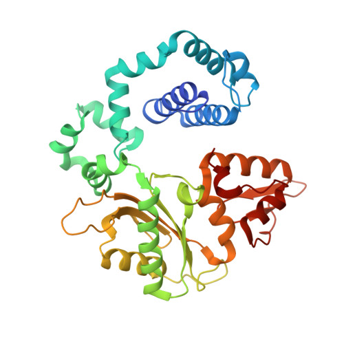
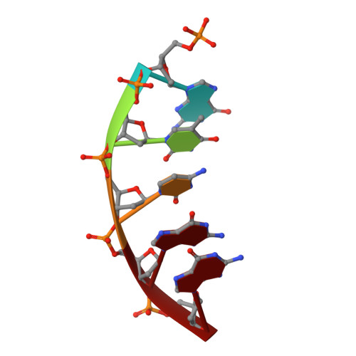
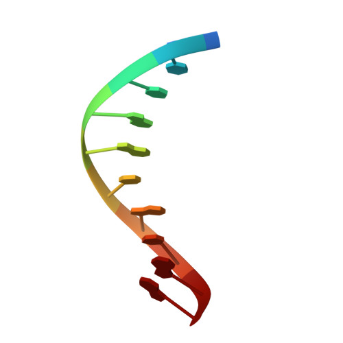
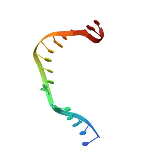
Structural Dynamics of a Common Mutagenic Oxidative DNA Lesion in Duplex DNA and during DNA Replication.

Ryan, B.J.Yang, H.Bacurio, J.H.T.Smith, M.R.Basu, A.K.Greenberg, M.M.Freudenthal, B.D.

(2022) J Am Chem Soc 144: 8054-8065

  • DOI: https://doi.org/10.1021/jacs.2c00193
  • Primary Citation of Related Structures:  
    7S9J, 7S9K, 7S9L, 7S9M, 7S9N, 7S9O, 7S9P, 7S9Q

  • PubMed Abstract: 

    N 6-(2-Deoxy-α,β-d- erythro -pentofuranosyl)-2,6-diamino-4-hydroxy-5-formamido pyrimidine (Fapy•dG) is a prevalent form of genomic DNA damage. Fapy•dG is formed in greater amounts under anoxic conditions than the well-studied, chemically related 7,8-dihydro-8-oxo-2'-deoxyguanosine (8-oxodGuo). Fapy•dG is more mutagenic in mammalian cells than 8-oxodGuo. A distinctive property of Fapy•dG is facile epimerization, but prior works with Fapy•dG analogues have precluded determining its effect on chemistry. We present crystallographic characterization of natural Fapy•dG in duplex DNA and as the template base for DNA polymerase β (Pol β). Fapy•dG adopts the β-anomer when base paired with cytosine but exists as a mixture of α- and β-anomers when promutagenically base paired with adenine. Rotation about the bond between the glycosidic nitrogen atom and the pyrimidine ring is also affected by the opposing nucleotide. Sodium cyanoborohydride soaking experiments trap the ring-opened Fapy•dG, demonstrating that ring opening and epimerization occur in the crystalline state. Ring opening and epimerization are facilitated by propitious water molecules that are observed in the structures. Determination of Fapy•dG mutagenicity in wild type and Pol β knockdown HEK 293T cells indicates that Pol β contributes to G → T transversions but also suppresses G → A transitions. Complementary kinetic studies have determined that Fapy•dG promotes mutagenesis by decreasing the catalytic efficiency of dCMP insertion opposite Fapy•dG, thus reducing polymerase fidelity. Kinetic studies have determined that dCMP incorporation opposite the β-anomer is ∼90 times faster than the α-anomer. This research identifies the importance of anomer dynamics, a feature unique to formamidopyrimidines, when considering the incorporation of nucleotides opposite Fapy•dG and potentially the repair of this structurally unusual lesion.


  • Organizational Affiliation

    Department of Biochemistry and Molecular Biology, and Department of Cancer Biology, University of Kansas Medical Center, Kansas City, Kansas 66160, United States.


Macromolecules

Find similar proteins by:  (by identity cutoff)  |  3D Structure
Entity ID: 1
MoleculeChains Sequence LengthOrganismDetailsImage
DNA polymerase beta335Homo sapiensMutation(s): 0 
Gene Names: POLB
EC: 2.7.7.7 (PDB Primary Data), 4.2.99 (PDB Primary Data)
UniProt & NIH Common Fund Data Resources
Find proteins for P06746 (Homo sapiens)
Explore P06746 
Go to UniProtKB:  P06746
PHAROS:  P06746
GTEx:  ENSG00000070501 
Entity Groups  
Sequence Clusters30% Identity50% Identity70% Identity90% Identity95% Identity100% Identity
UniProt GroupP06746
Sequence Annotations
Expand
  • Reference Sequence

Find similar nucleic acids by:  Sequence   |   3D Structure  

Entity ID: 2
MoleculeChains LengthOrganismImage
DNA (5'-D(P*GP*TP*CP*GP*G)-3')B [auth D]5synthetic construct
Sequence Annotations
Expand
  • Reference Sequence

Find similar nucleic acids by:  Sequence   |   3D Structure  

Entity ID: 3
MoleculeChains LengthOrganismImage
DNA (5'-D(*GP*CP*TP*GP*AP*TP*GP*CP*GP*A)-3')C [auth P]10synthetic construct
Sequence Annotations
Expand
  • Reference Sequence

Find similar nucleic acids by:  Sequence   |   3D Structure  

Entity ID: 4
MoleculeChains LengthOrganismImage
DNA (5'-D(*CP*CP*GP*AP*CP*(FAP)P*TP*CP*GP*CP*AP*TP*CP*AP*GP*C)-3')D [auth T]16synthetic construct
Sequence Annotations
Expand
  • Reference Sequence
Small Molecules
Ligands 4 Unique
IDChains Name / Formula / InChI Key2D Diagram3D Interactions
F2A
Query on F2A

Download Ideal Coordinates CCD File 
J [auth A]2'-deoxy-5'-O-[(S)-hydroxy{[(S)-hydroxy(phosphonooxy)phosphoryl]methyl}phosphoryl]adenosine
C11 H18 N5 O11 P3
XETARULVTCYJAN-XLPZGREQSA-N
MN
Query on MN

Download Ideal Coordinates CCD File 
G [auth A],
H [auth A],
K [auth A]
MANGANESE (II) ION
Mn
WAEMQWOKJMHJLA-UHFFFAOYSA-N
MG
Query on MG

Download Ideal Coordinates CCD File 
L [auth A],
M [auth A]
MAGNESIUM ION
Mg
JLVVSXFLKOJNIY-UHFFFAOYSA-N
NA
Query on NA

Download Ideal Coordinates CCD File 
E [auth A],
F [auth A],
I [auth A]
SODIUM ION
Na
FKNQFGJONOIPTF-UHFFFAOYSA-N
Experimental Data & Validation

Experimental Data

  • Method: X-RAY DIFFRACTION
  • Resolution: 1.90 Å
  • R-Value Free: 0.247 
  • R-Value Work: 0.193 
  • R-Value Observed: 0.197 
  • Space Group: P 1 21 1
Unit Cell:
Length ( Å )Angle ( ˚ )
a = 54.624α = 90
b = 78.583β = 112.435
c = 54.916γ = 90
Software Package:
Software NamePurpose
PHENIXrefinement
PHENIXphasing
HKL-2000data reduction
HKL-2000data scaling
HKL-2000data collection
PDB_EXTRACTdata extraction

Structure Validation

View Full Validation Report


Ligand Structure Quality Assessment 


Entry History & Funding Information

Deposition Data


Funding OrganizationLocationGrant Number
National Institutes of Health/National Institute of Environmental Health Sciences (NIH/NIEHS)United States--

Revision History  (Full details and data files)

  • Version 1.0: 2022-08-03
    Type: Initial release
  • Version 1.1: 2023-10-18
    Changes: Data collection, Refinement description