7TJT

Yeast ATP synthase F1 region State 1-3catalytic beta_tight open without exogenous ATP

  • Classification: HYDROLASE
  • Organism(s): Saccharomyces cerevisiae
  • Mutation(s): No 

  • Deposited: 2022-01-17 Released: 2022-04-20 
  • Deposition Author(s): Guo, H., Rubinstein, J.L.
  • Funding Organization(s): Canada Research Chairs, Canadian Institutes of Health Research (CIHR), Canada Foundation for Innovation

Experimental Data Snapshot

  • Method: ELECTRON MICROSCOPY
  • Resolution: 3.20 Å
  • Aggregation State: PARTICLE 
  • Reconstruction Method: SINGLE PARTICLE 

wwPDB Validation   3D Report Full Report


This is version 1.2 of the entry. See complete history


Literature

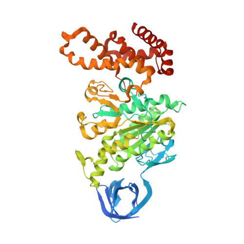
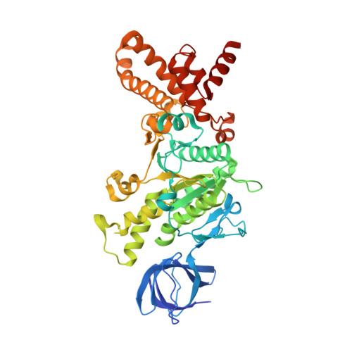
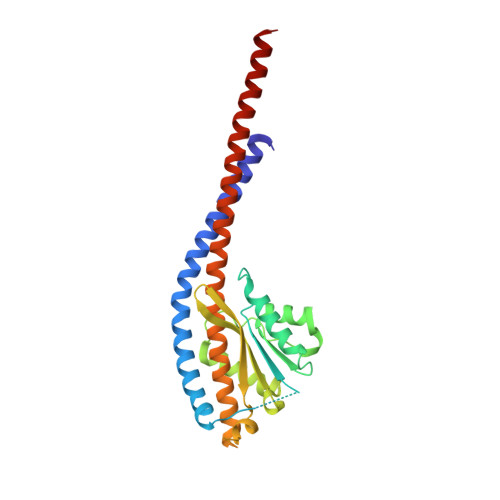
Structure of ATP synthase under strain during catalysis.

Guo, H.Rubinstein, J.L.

(2022) Nat Commun 13: 2232-2232

  • DOI: https://doi.org/10.1038/s41467-022-29893-2
  • Primary Citation of Related Structures:  
    7TJS, 7TJT, 7TJU, 7TJV, 7TJW, 7TJX, 7TJY, 7TJZ, 7TK0, 7TK1, 7TK2, 7TK3, 7TK4, 7TK5, 7TK6, 7TK7, 7TK8, 7TK9, 7TKA, 7TKB, 7TKC, 7TKD, 7TKE, 7TKF, 7TKG, 7TKH, 7TKI, 7TKJ, 7TKK, 7TKL, 7TKM, 7TKN, 7TKO, 7TKP, 7TKQ, 7TKR, 7TKS

  • PubMed Abstract: 

    ATP synthases are macromolecular machines consisting of an ATP-hydrolysis-driven F 1 motor and a proton-translocation-driven F O motor. The F 1 and F O motors oppose each other's action on a shared rotor subcomplex and are held stationary relative to each other by a peripheral stalk. Structures of resting mitochondrial ATP synthases revealed a left-handed curvature of the peripheral stalk even though rotation of the rotor, driven by either ATP hydrolysis in F 1 or proton translocation through F O , would apply a right-handed bending force to the stalk. We used cryoEM to image yeast mitochondrial ATP synthase under strain during ATP-hydrolysis-driven rotary catalysis, revealing a large deformation of the peripheral stalk. The structures show how the peripheral stalk opposes the bending force and suggests that during ATP synthesis proton translocation causes accumulation of strain in the stalk, which relaxes by driving the relative rotation of the rotor through six sub-steps within F 1 , leading to catalysis.


  • Organizational Affiliation

    Molecular Medicine Program, The Hospital for Sick Children, Toronto, Ontario, Canada.


Macromolecules
Find similar proteins by:  (by identity cutoff)  |  3D Structure
Entity ID: 1
MoleculeChains Sequence LengthOrganismDetailsImage
ATP synthase subunit alpha
A, B, C
510Saccharomyces cerevisiaeMutation(s): 0 
UniProt
Find proteins for P07251 (Saccharomyces cerevisiae (strain ATCC 204508 / S288c))
Explore P07251 
Go to UniProtKB:  P07251
Entity Groups  
Sequence Clusters30% Identity50% Identity70% Identity90% Identity95% Identity100% Identity
UniProt GroupP07251
Sequence Annotations
Expand
  • Reference Sequence
Find similar proteins by:  (by identity cutoff)  |  3D Structure
Entity ID: 2
MoleculeChains Sequence LengthOrganismDetailsImage
ATP synthase subunit beta
D, E, F
478Saccharomyces cerevisiaeMutation(s): 0 
EC: 7.1.2.2
UniProt
Find proteins for P00830 (Saccharomyces cerevisiae (strain ATCC 204508 / S288c))
Explore P00830 
Go to UniProtKB:  P00830
Entity Groups  
Sequence Clusters30% Identity50% Identity70% Identity90% Identity95% Identity100% Identity
UniProt GroupP00830
Sequence Annotations
Expand
  • Reference Sequence
Find similar proteins by:  (by identity cutoff)  |  3D Structure
Entity ID: 3
MoleculeChains Sequence LengthOrganismDetailsImage
ATP synthase subunit gamma278Saccharomyces cerevisiaeMutation(s): 0 
UniProt
Find proteins for P38077 (Saccharomyces cerevisiae (strain ATCC 204508 / S288c))
Explore P38077 
Go to UniProtKB:  P38077
Entity Groups  
Sequence Clusters30% Identity50% Identity70% Identity90% Identity95% Identity100% Identity
UniProt GroupP38077
Sequence Annotations
Expand
  • Reference Sequence
Experimental Data & Validation

Experimental Data

  • Method: ELECTRON MICROSCOPY
  • Resolution: 3.20 Å
  • Aggregation State: PARTICLE 
  • Reconstruction Method: SINGLE PARTICLE 
EM Software:
TaskSoftware PackageVersion
MODEL REFINEMENTCoot0.9.2
MODEL REFINEMENTISOLDE1.2.0
MODEL REFINEMENTPHENIX1.19.2
RECONSTRUCTIONcryoSPARC3.1.0

Structure Validation

View Full Validation Report


Entry History & Funding Information

Deposition Data


Funding OrganizationLocationGrant Number
Canada Research ChairsCanadaElectron Cryomicroscopy
Canadian Institutes of Health Research (CIHR)CanadaPJT162186
Canada Foundation for InnovationCanada--

Revision History  (Full details and data files)

  • Version 1.0: 2022-04-20
    Type: Initial release
  • Version 1.1: 2022-05-11
    Changes: Database references
  • Version 1.2: 2024-02-21
    Changes: Data collection, Refinement description