9H49

Crystal structure of the adduct between human serum transferrin (apo-form) and cisplatin


Experimental Data Snapshot

  • Method: X-RAY DIFFRACTION
  • Resolution: 3.52 Å
  • R-Value Free: 
    0.249 (Depositor), 0.250 (DCC) 
  • R-Value Work: 
    0.231 (Depositor), 0.231 (DCC) 
  • R-Value Observed: 
    0.232 (Depositor) 

Starting Model: experimental
View more details

wwPDB Validation   3D Report Full Report


This is version 1.1 of the entry. See complete history


Literature

Cisplatin/Apo-Transferrin Adduct: X-ray Structure and Binding to the Transferrin Receptor 1.

Troisi, R.Galardo, F.Ferraro, G.Lucignano, R.Picone, D.Marano, A.Trifuoggi, M.Sica, F.Merlino, A.

(2025) Inorg Chem 64: 761-765

  • DOI: https://doi.org/10.1021/acs.inorgchem.4c04435
  • Primary Citation of Related Structures:  
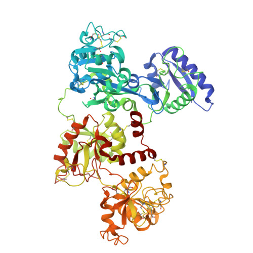
    9H49

  • PubMed Abstract: 

    Here, we report the X-ray structure of the adduct formed upon reaction of cisplatin, one of the most prescribed anticancer agents for the clinic treatment of solid tumors, with the apo-form of human serum transferrin (hTF). Two Pt binding sites were identified in both molecules of the adduct present in the crystal asymmetric unit: Pt binds close to the side chains of Met256 and Met499 at the N- and C-lobe, respectively. In the crystal structure, the cisplatin moiety bound to Met256 also interacts with Ser616 from a symmetry related molecule. Structural analyses, together with in solution data, demonstrate that the presence of iron does not affect the ability of hTF to bind cisplatin and that the cisplatin binding does not significantly alter the overall conformation of the different forms of the protein that remain able to form a complex with the transferrin receptor 1 (TfR1). These data suggest that the different hTF forms can be used as nanocarriers for targeted (combined) metallodrug delivery.


  • Organizational Affiliation

    Department of Chemical Sciences, University of Naples Federico II, Complesso Universitario di Monte Sant'Angelo, via Cintia, I-80126, Naples, Italy.


Macromolecules
Find similar proteins by:  (by identity cutoff)  |  3D Structure
Entity ID: 1
MoleculeChains Sequence LengthOrganismDetailsImage
Serotransferrin
A, B
679Homo sapiensMutation(s): 0 
UniProt & NIH Common Fund Data Resources
Find proteins for P02787 (Homo sapiens)
Explore P02787 
Go to UniProtKB:  P02787
PHAROS:  P02787
GTEx:  ENSG00000091513 
Entity Groups  
Sequence Clusters30% Identity50% Identity70% Identity90% Identity95% Identity100% Identity
UniProt GroupP02787
Sequence Annotations
Expand
  • Reference Sequence
Small Molecules
Ligands 3 Unique
IDChains Name / Formula / InChI Key2D Diagram3D Interactions
PT (Subject of Investigation/LOI)
Query on PT

Download Ideal Coordinates CCD File 
F [auth A],
I [auth A],
O [auth B],
R [auth B]
PLATINUM (II) ION
Pt
HRGDZIGMBDGFTC-UHFFFAOYSA-N
CIT
Query on CIT

Download Ideal Coordinates CCD File 
C [auth A]
D [auth A]
E [auth A]
L [auth B]
M [auth B]
CITRIC ACID
C6 H8 O7
KRKNYBCHXYNGOX-UHFFFAOYSA-N
NH3
Query on NH3

Download Ideal Coordinates CCD File 
G [auth A]
H [auth A]
J [auth A]
K [auth A]
P [auth B]
AMMONIA
H3 N
QGZKDVFQNNGYKY-UHFFFAOYSA-N
Experimental Data & Validation

Experimental Data

  • Method: X-RAY DIFFRACTION
  • Resolution: 3.52 Å
  • R-Value Free:  0.249 (Depositor), 0.250 (DCC) 
  • R-Value Work:  0.231 (Depositor), 0.231 (DCC) 
  • R-Value Observed: 0.232 (Depositor) 
Space Group: P 21 21 21
Unit Cell:
Length ( Å )Angle ( ˚ )
a = 88.184α = 90
b = 103.68β = 90
c = 200.283γ = 90
Software Package:
Software NamePurpose
REFMACrefinement
XDSdata reduction
Aimlessdata scaling
PHASERphasing
autoPROCdata processing

Structure Validation

View Full Validation Report


Entry History & Funding Information

Deposition Data


Funding OrganizationLocationGrant Number
Ministero dell Universita e della RicercaItaly2022JMFC3X

Revision History  (Full details and data files)

  • Version 1.0: 2025-01-22
    Type: Initial release
  • Version 1.1: 2025-01-29
    Changes: Database references