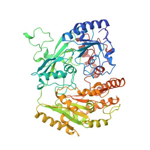
Crystal structures and inhibitor binding in the octameric flavoenzyme vanillyl-alcohol oxidase: the shape of the active-site cavity controls substrate specificity.
Mattevi, A., Fraaije, M.W., Mozzarelli, A., Olivi, L., Coda, A., van Berkel, W.J.(1997) Structure 5: 907-920
- PubMed: 9261083
- DOI: https://doi.org/10.1016/s0969-2126(97)00245-1
- Primary Citation of Related Structures:
1AHU, 1AHV, 1AHZ, 1VAO, 2VAO - PubMed Abstract:
Lignin degradation leads to the formation of a broad spectrum of aromatic molecules that can be used by various fungal micro-organisms as their sole source of carbon. When grown on phenolic compounds, Penicillium simplicissimum induces the strong impression of a flavin-containing vanillyl-alcohol oxidase (VAO). The enzyme catalyses the oxidation of a vast array of substrates, ranging from aromatic amines to 4-alkyphenols. VAO is a member of a novel class of widely distributed oxidoreductases, which use flavin adenine dinucleotide (FAD) as a cofactor covalently bound to the protein. We have carried out the determination of the structure of VAO in order to shed light on the most interesting features of these novel oxidoreductases, such as the functional significance of covalent flavinylation and the mechanism of catalysis. The crystal structure of VAO has been determined in the native state and in complexes with four inhibitors. The enzyme is an octamer with 42 symmetry; the inhibitors bind in a hydrophobic, elongated cavity on the si side of the flavin molecule. Three residues, Tyr108, Tyr503 and Arg504 form an anion-binding subsite, which stabilises the phenolate form of the substrate. The structure of VAO complexed with the inhibitor 4-(1-heptenyl)phenol shows that the catalytic cavity is completely filled by the inhibitor, explaining why alkylphenols bearing aliphatic substituents longer than seven carbon atoms do not bind to the enzyme. The shape of the active-site cavity controls substrate specificity by providing a 'size exclusion mechanism'. Inside the cavity, the substrate aromatic ring is positioned at an angle of 18 degrees to the flavin ring. This arrangement is ideally suited for a hydride transfer reaction, which is further facilitated by substrate deprotonation. Burying the substrate beneath the protein surface is a recurrent strategy, common to many flavoenzymes that effect substrate oxidation or reduction via hydride transfer.
Organizational Affiliation:
Department of Genetics & Microbiology, University of Pavia, Italy. MATTEVI@IPVGEN.UNIPV.IT