1CM4

Motions of calmodulin-four-conformer refinement


Experimental Data Snapshot

  • Method: X-RAY DIFFRACTION
  • Resolution: 2.00 Å
  • R-Value Free: 0.262 
  • R-Value Work: 0.166 
  • R-Value Observed: 0.166 

wwPDB Validation   3D Report Full Report


This is version 1.4 of the entry. See complete history


Literature

Motions of calmodulin characterized using both Bragg and diffuse X-ray scattering.

Wall, M.E.Clarage, J.B.Phillips Jr., G.N.

(1997) Structure 5: 1599-1612

  • DOI: https://doi.org/10.1016/s0969-2126(97)00308-0
  • Primary Citation of Related Structures:  
    1CM1, 1CM4

  • PubMed Abstract: 

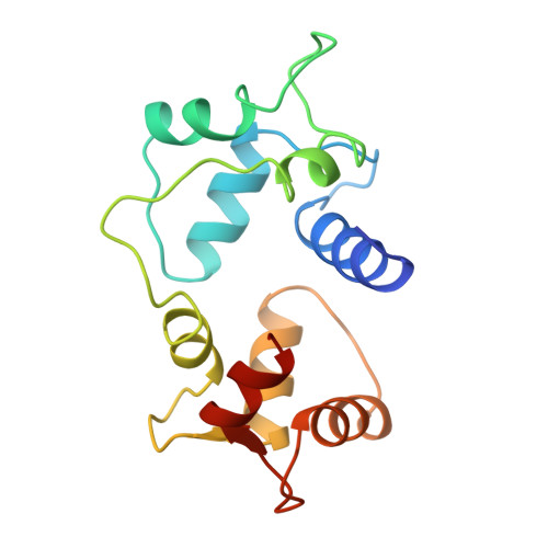
    Calmodulin is a calcium-activated regulatory protein which can bind to many different targets. The protein resembles a highly flexible dumbbell, and bends in the middle as it binds. This and other motions must be understood to formulate a realistic model of calmodulin function. Using the Bragg reflections from X-ray crystallography, a multiple-conformer refinement of a calmodulin-peptide complex shows anisotropic displacements, with high variations of dihedral angles in several nonhelical domains: the flexible linker; three of the four calcium-binding sites (including both of the N-terminal sites); and a turn connecting the C-terminal EF-hand calcium-binding domains. Three-dimensional maps of the large scale diffuse X-ray scattering data show isotropic liquid-like motions with an unusually small correlation length. Three-dimensional maps of the small scale diffuse streaks show highly coupled, anisotropic motions along the head-to-tail molecular packing direction in the unit cell. There is also weak coupling perpendicular to the head-to-tail packing direction, particularly across a cavity occupied by the disordered linker domain of the molecule. Together, the Bragg and diffuse scattering present a self-consistent description of the motions in the flexible linker of calmodulin. The other mobile regions of the protein are also of great interest. In particular, the high variations in the calcium-binding sites are likely to influence how strongly they bind ions. This is especially important in the N-terminal sites, which regulate the activity of the molecule.


  • Organizational Affiliation

    Department of Biochemistry and Cell Biology, The WM Keck Center for Computational Biology, Rice University Houston, TX 77005-1892, USA, mewall@bioc.rice.edu


Macromolecules
Find similar proteins by:  (by identity cutoff)  |  3D Structure
Entity ID: 1
MoleculeChains Sequence LengthOrganismDetailsImage
CALMODULIN148Bos taurusMutation(s): 0 
UniProt
Find proteins for P62157 (Bos taurus)
Explore P62157 
Go to UniProtKB:  P62157
Entity Groups  
Sequence Clusters30% Identity50% Identity70% Identity90% Identity95% Identity100% Identity
UniProt GroupP62157
Sequence Annotations
Expand
  • Reference Sequence

Find similar proteins by:  Sequence   |   3D Structure  

Entity ID: 2
MoleculeChains Sequence LengthOrganismDetailsImage
CALMODULIN-DEPENDENT PROTEIN KINASE II-ALPHA25Rattus norvegicusMutation(s): 0 
Gene Names: Camk2a
EC: 2.7.1.123
UniProt
Find proteins for P11275 (Rattus norvegicus)
Explore P11275 
Go to UniProtKB:  P11275
Entity Groups  
Sequence Clusters30% Identity50% Identity70% Identity90% Identity95% Identity100% Identity
UniProt GroupP11275
Sequence Annotations
Expand
  • Reference Sequence
Small Molecules
Ligands 1 Unique
IDChains Name / Formula / InChI Key2D Diagram3D Interactions
CA
Query on CA

Download Ideal Coordinates CCD File 
C [auth A]
D [auth A]
E [auth A]
F [auth A]
G [auth A]
C [auth A],
D [auth A],
E [auth A],
F [auth A],
G [auth A],
H [auth A],
I [auth A],
J [auth A],
K [auth A],
L [auth A],
M [auth A],
N [auth A],
O [auth A],
P [auth A],
Q [auth A],
R [auth A],
S [auth A]
CALCIUM ION
Ca
BHPQYMZQTOCNFJ-UHFFFAOYSA-N
Experimental Data & Validation

Experimental Data

  • Method: X-RAY DIFFRACTION
  • Resolution: 2.00 Å
  • R-Value Free: 0.262 
  • R-Value Work: 0.166 
  • R-Value Observed: 0.166 
  • Space Group: C 2 2 21
Unit Cell:
Length ( Å )Angle ( ˚ )
a = 38.751α = 90
b = 75.209β = 90
c = 120.073γ = 90
Software Package:
Software NamePurpose
X-PLORmodel building
X-PLORrefinement
DENZOdata reduction
SCALEPACKdata scaling
X-PLORphasing

Structure Validation

View Full Validation Report


Entry History 

Deposition Data

Revision History  (Full details and data files)

  • Version 1.0: 1998-03-04
    Type: Initial release
  • Version 1.1: 2008-03-24
    Changes: Version format compliance
  • Version 1.2: 2011-07-13
    Changes: Derived calculations, Version format compliance
  • Version 1.3: 2013-07-24
    Changes: Structure summary
  • Version 1.4: 2023-08-09
    Changes: Database references, Derived calculations, Other, Refinement description