1KMZ

MOLECULAR BASIS OF MITOMYCIN C RESICTANCE IN STREPTOMYCES: CRYSTAL STRUCTURES OF THE MRD PROTEIN WITH AND WITHOUT A DRUG DERIVATIVE


Experimental Data Snapshot

  • Method: X-RAY DIFFRACTION
  • Resolution: 1.50 Å
  • R-Value Free: 0.231 
  • R-Value Observed: 0.163 

wwPDB Validation   3D Report Full Report


This is version 1.3 of the entry. See complete history


Literature

Molecular basis of mitomycin C resistance in streptomyces: structure and function of the MRD protein.

Martin, T.W.Dauter, Z.Devedjiev, Y.Sheffield, P.Jelen, F.He, M.Sherman, D.H.Otlewski, J.Derewenda, Z.S.Derewenda, U.

(2002) Structure 10: 933-942

  • DOI: https://doi.org/10.1016/s0969-2126(02)00778-5
  • Primary Citation of Related Structures:  
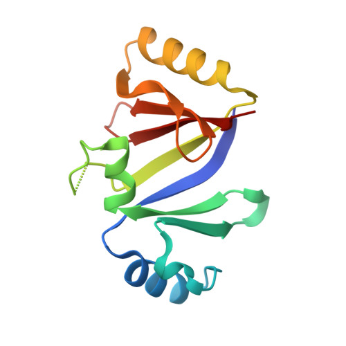
    1KLL, 1KMZ

  • PubMed Abstract: 

    Mitomycin C (MC) is a potent anticancer agent. Streptomyces lavendulae, which produces MC, protects itself from the lethal effects of the drug by expressing several resistance proteins. One of them (MRD) binds MC and functions as a drug exporter. We report the crystal structure of MRD and its complex with an MC metabolite, 1,2-cis-1-hydroxy-2,7-diaminomitosene, at 1.5 A resolution. The drug is sandwiched by pi-stacking interactions of His-38 and Trp-108. MRD is a dimer. The betaalphabetabetabeta fold of the MRD molecule is reminiscent of methylmalonyl-CoA epimerase, bleomycin resistance proteins, glyoxalase I, and extradiol dioxygenases. The location of the binding site is identical to the ones in evolutionarily related enzymes, suggesting that the protein may have been recruited from a different metabolic pathway.


  • Organizational Affiliation

    Department of Molecular Physiology and Biological Physics, University of Virginia, Health Sciences System, Charlottesville 22908, USA.


Macromolecules
Find similar proteins by:  (by identity cutoff)  |  3D Structure
Entity ID: 1
MoleculeChains Sequence LengthOrganismDetailsImage
mitomycin-binding protein130Streptomyces lavendulaeMutation(s): 0 
UniProt
Find proteins for O05205 (Streptomyces lavendulae)
Explore O05205 
Go to UniProtKB:  O05205
Entity Groups  
Sequence Clusters30% Identity50% Identity70% Identity90% Identity95% Identity100% Identity
UniProt GroupO05205
Sequence Annotations
Expand
  • Reference Sequence
Experimental Data & Validation

Experimental Data

  • Method: X-RAY DIFFRACTION
  • Resolution: 1.50 Å
  • R-Value Free: 0.231 
  • R-Value Observed: 0.163 
  • Space Group: C 1 2 1
Unit Cell:
Length ( Å )Angle ( ˚ )
a = 44.508α = 90
b = 60.651β = 105.97
c = 43.776γ = 90
Software Package:
Software NamePurpose
SHELXmodel building
SHELXL-97refinement
DENZOdata reduction
SCALEPACKdata scaling
SHELXphasing

Structure Validation

View Full Validation Report


Entry History 

Deposition Data

Revision History  (Full details and data files)

  • Version 1.0: 2002-07-19
    Type: Initial release
  • Version 1.1: 2008-04-27
    Changes: Version format compliance
  • Version 1.2: 2011-07-13
    Changes: Derived calculations, Version format compliance
  • Version 1.3: 2023-08-16
    Changes: Data collection, Database references, Refinement description