1XNZ

Crystal Structure of Mn(II) form of E. coli. Methionine Aminopeptidase in complex with 5-(2-chlorophenyl)furan-2-carboxylic acid


Experimental Data Snapshot

  • Method: X-RAY DIFFRACTION
  • Resolution: 1.52 Å
  • R-Value Free: 0.250 
  • R-Value Work: 0.219 
  • R-Value Observed: 0.244 

wwPDB Validation   3D Report Full Report


Ligand Structure Quality Assessment 


This is version 1.3 of the entry. See complete history


Literature

Metalloform-Selective Inhibitors of Escherichia coli Methionine Aminopeptidase and X-ray Structure of a Mn(II)-Form Enzyme Complexed with an Inhibitor.

Ye, Q.-Z.Xie, S.-X.Huang, M.Huang, W.-J.Lu, J.-P.Ma, Z.-Q.

(2004) J Am Chem Soc 126: 13940-13941

  • DOI: https://doi.org/10.1021/ja045864p
  • Primary Citation of Related Structures:  
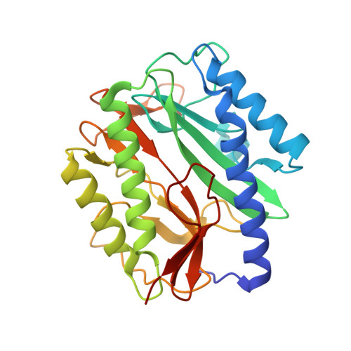
    1XNZ

  • PubMed Abstract: 

    Methionine aminopeptidase (MetAP) enzymes require a divalent metal ion such as Mn(II), Fe(II), Co(II), Ni(II), or Zn(II) for its removal of the N-terminal methionine from newly synthesized proteins, but it is not certain which of these ions is most important in vivo. Metalloform-selective MetAP inhibitors could be valuable for defining which metals are physiologically relevant for MetAP activation and could serve as leads for development of new therapeutic agents. We have screened a library of 43 736 small drug-like molecules against Escherichia coli MetAP and identified two groups of potent and highly metalloform-selective inhibitors of the Co(II)-form, and of the Mn(II)-form, of this enzyme. Compound 1 is 790-fold more selective for the Co(II)-form, while compound 4 is over 640-fold more potent toward the Mn(II)-form. The X-ray structure of a di-Mn(II) form of E. coli MetAP complexed with the Mn(II)-form-selective compound 4 was obtained, and it shows that the inhibitor interacts with both Mn(II) ions through the two oxygen atoms of its free carboxylate group. The preferential coordination of the hard (oxygen) donors to Mn(II) may contribute to its superb selectivity toward the Mn(II)-form.


  • Organizational Affiliation

    High Throughput Screening Laboratory and Protein Structure Laboratory, University of Kansas, Lawrence, Kansas 66045, USA. qye@ku.edu


Macromolecules
Find similar proteins by:  (by identity cutoff)  |  3D Structure
Entity ID: 1
MoleculeChains Sequence LengthOrganismDetailsImage
Methionine aminopeptidase264Escherichia coliMutation(s): 0 
Gene Names: map
EC: 3.4.11.18
UniProt
Find proteins for P0AE18 (Escherichia coli (strain K12))
Explore P0AE18 
Go to UniProtKB:  P0AE18
Entity Groups  
Sequence Clusters30% Identity50% Identity70% Identity90% Identity95% Identity100% Identity
UniProt GroupP0AE18
Sequence Annotations
Expand
  • Reference Sequence
Small Molecules
Binding Affinity Annotations 
IDSourceBinding Affinity
FCD BindingDB:  1XNZ IC50: min: 510, max: 2.00e+5 (nM) from 7 assay(s)
Binding MOAD:  1XNZ IC50: 240 (nM) from 1 assay(s)
PDBBind:  1XNZ IC50: 240 (nM) from 1 assay(s)
Experimental Data & Validation

Experimental Data

  • Method: X-RAY DIFFRACTION
  • Resolution: 1.52 Å
  • R-Value Free: 0.250 
  • R-Value Work: 0.219 
  • R-Value Observed: 0.244 
  • Space Group: P 1 21 1
Unit Cell:
Length ( Å )Angle ( ˚ )
a = 37.6α = 90
b = 60.3β = 104.8
c = 49.7γ = 90
Software Package:
Software NamePurpose
CrystalCleardata collection
XDSdata reduction
AMoREphasing
CNSrefinement
CrystalCleardata reduction
XDSdata scaling

Structure Validation

View Full Validation Report


Ligand Structure Quality Assessment 


Entry History 

Deposition Data

Revision History  (Full details and data files)

  • Version 1.0: 2004-11-02
    Type: Initial release
  • Version 1.1: 2008-04-30
    Changes: Version format compliance
  • Version 1.2: 2011-07-13
    Changes: Version format compliance
  • Version 1.3: 2024-02-14
    Changes: Data collection, Database references, Derived calculations