5Y6U

Crystal structure of wild-type YabJ protein from Bacillus subtilis (natto).


Experimental Data Snapshot

  • Method: X-RAY DIFFRACTION
  • Resolution: 1.50 Å
  • R-Value Free: 0.180 
  • R-Value Work: 0.161 
  • R-Value Observed: 0.162 

wwPDB Validation   3D Report Full Report


This is version 1.2 of the entry. See complete history


Literature

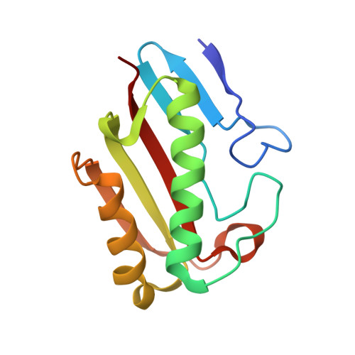
Tetramer formation of Bacillus subtilis YabJ protein that belongs to YjgF/YER057c/UK114 family.

Fujimoto, Z.Hong, L.T.T.Kishine, N.Suzuki, N.Kimura, K.

(2021) Biosci Biotechnol Biochem 85: 297-306

  • DOI: https://doi.org/10.1093/bbb/zbaa037
  • Primary Citation of Related Structures:  
    5Y6U, 7CD2, 7CD3, 7CD4

  • PubMed Abstract: 

    Bacillus subtilis YabJ protein belongs to the highly conserved YjgF/YER057c/UK114 family, which has a homotrimeric quaternary structure. The dominant allele of yabJ gene that is caused by a single amino acid mutation of Ser103Phe enables poly-γ-glutamic acid (γPGA) production of B. subtilis under conditions where the cell-density signal transduction was disturbed by the loss of DegQ function. X-ray crystallography of recombinant proteins revealed that unlike the homotrimeric wild-type YabJ, the mutant YabJ(Ser103Phe) had a homotetrameric quaternary structure, and the structural change appeared to be triggered by an inversion of the fifth β-strand. The YabJ homotetramer has a hole that is highly accessible, penetrating through the tetramer, and 2 surface concaves as potential ligand-binding sites. Western blot analyses revealed that the conformational change was also induced in vivo by the Ser103Phe mutation.


  • Organizational Affiliation

    Advanced Analysis Center, National Agriculture and Food Research Organization (NAAC/NARO), Tsukuba, Ibaraki, Japan.


Macromolecules
Find similar proteins by:  (by identity cutoff)  |  3D Structure
Entity ID: 1
MoleculeChains Sequence LengthOrganismDetailsImage
YabJ protein
A, B, C
125Bacillus subtilis subsp. natto BEST195Mutation(s): 0 
Gene Names: yabJBSNT_00084
UniProt
Find proteins for D4G3D4 (Bacillus subtilis subsp. natto (strain BEST195))
Explore D4G3D4 
Go to UniProtKB:  D4G3D4
Entity Groups  
Sequence Clusters30% Identity50% Identity70% Identity90% Identity95% Identity100% Identity
UniProt GroupD4G3D4
Sequence Annotations
Expand
  • Reference Sequence
Experimental Data & Validation

Experimental Data

  • Method: X-RAY DIFFRACTION
  • Resolution: 1.50 Å
  • R-Value Free: 0.180 
  • R-Value Work: 0.161 
  • R-Value Observed: 0.162 
  • Space Group: P 65
Unit Cell:
Length ( Å )Angle ( ˚ )
a = 53.767α = 90
b = 53.767β = 90
c = 203.941γ = 120
Software Package:
Software NamePurpose
REFMACrefinement
HKL-2000data reduction
SCALEPACKdata scaling
MOLREPphasing

Structure Validation

View Full Validation Report


Entry History 

Deposition Data

Revision History  (Full details and data files)

  • Version 1.0: 2018-08-15
    Type: Initial release
  • Version 1.1: 2021-03-03
    Changes: Database references
  • Version 1.2: 2023-11-22
    Changes: Data collection, Database references, Refinement description