6GZC

heterotetrameric katanin p60:p80 complex


Experimental Data Snapshot

  • Method: X-RAY DIFFRACTION
  • Resolution: 2.00 Å
  • R-Value Free: 0.228 
  • R-Value Work: 0.209 
  • R-Value Observed: 0.210 

wwPDB Validation   3D Report Full Report


This is version 1.2 of the entry. See complete history


Literature

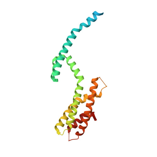
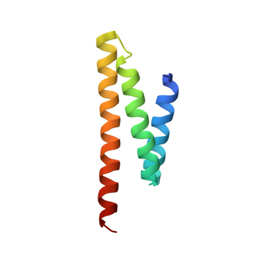
Crystal Structure of a Heterotetrameric Katanin p60:p80 Complex.

Faltova, L.Jiang, K.Frey, D.Wu, Y.Capitani, G.Prota, A.E.Akhmanova, A.Steinmetz, M.O.Kammerer, R.A.

(2019) Structure 27: 1375-1383.e3

  • DOI: https://doi.org/10.1016/j.str.2019.07.002
  • Primary Citation of Related Structures:  
    6GZC

  • PubMed Abstract: 

    Katanin is a microtubule-severing enzyme that is crucial for many cellular processes. Katanin consists of two subunits, p60 and p80, that form a stable complex. The interaction between subunits is mediated by the p60 N-terminal microtubule-interacting and -trafficking domain (p60-MIT) and the p80 C-terminal domain (p80-CTD). Here, we performed a biophysical characterization of the mouse p60-MIT:p80-CTD heterodimer and show that this complex can assemble into heterotetramers. We identified two mutations that enhance heterotetramer formation and determined the X-ray crystal structure of this mutant complex. The structure revealed a domain-swapped heterotetramer consisting of two p60-MIT:p80-CTD heterodimers. Structure-based sequence alignments suggest that heterotetramerization of katanin might be a common feature of various species. Furthermore, we show that enhanced heterotetramerization of katanin impairs its microtubule end-binding properties and increases the enzyme's microtubule lattice binding and severing activities. Therefore, our findings suggest the existence of different katanin oligomers that possess distinct functional properties.


  • Organizational Affiliation

    Laboratory of Biomolecular Research, Division of Biology and Chemistry, Paul Scherrer Institut, 5232 Villigen PSI, Switzerland.


Macromolecules
Find similar proteins by:  (by identity cutoff)  |  3D Structure
Entity ID: 1
MoleculeChains Sequence LengthOrganismDetailsImage
Katanin p80 WD40 repeat-containing subunit B1A,
D [auth C]
212Mus musculusMutation(s): 0 
Gene Names: Katnb1
UniProt & NIH Common Fund Data Resources
Find proteins for Q8BG40 (Mus musculus)
Explore Q8BG40 
Go to UniProtKB:  Q8BG40
IMPC:  MGI:1921437
Entity Groups  
Sequence Clusters30% Identity50% Identity70% Identity90% Identity95% Identity100% Identity
UniProt GroupQ8BG40
Sequence Annotations
Expand
  • Reference Sequence
Find similar proteins by:  (by identity cutoff)  |  3D Structure
Entity ID: 2
MoleculeChains Sequence LengthOrganismDetailsImage
Katanin p60 ATPase-containing subunit A1B,
C [auth D]
80Mus musculusMutation(s): 0 
Gene Names: Katna1
EC: 3.6.4.3
UniProt & NIH Common Fund Data Resources
Find proteins for Q9WV86 (Mus musculus)
Explore Q9WV86 
Go to UniProtKB:  Q9WV86
IMPC:  MGI:1344353
Entity Groups  
Sequence Clusters30% Identity50% Identity70% Identity90% Identity95% Identity100% Identity
UniProt GroupQ9WV86
Sequence Annotations
Expand
  • Reference Sequence
Experimental Data & Validation

Experimental Data

  • Method: X-RAY DIFFRACTION
  • Resolution: 2.00 Å
  • R-Value Free: 0.228 
  • R-Value Work: 0.209 
  • R-Value Observed: 0.210 
  • Space Group: P 1
Unit Cell:
Length ( Å )Angle ( ˚ )
a = 45.49α = 108.32
b = 57.85β = 101
c = 59.77γ = 98.26
Software Package:
Software NamePurpose
PHENIXrefinement
XDSdata reduction
XSCALEdata scaling
PHASERphasing

Structure Validation

View Full Validation Report


Entry History 

Deposition Data

Revision History  (Full details and data files)

  • Version 1.0: 2019-08-07
    Type: Initial release
  • Version 1.1: 2019-09-18
    Changes: Data collection, Database references
  • Version 1.2: 2024-01-17
    Changes: Data collection, Database references, Refinement description