6HPP

X-ray structure of GLIC in complex with propionate


Experimental Data Snapshot

  • Method: X-RAY DIFFRACTION
  • Resolution: 3.20 Å
  • R-Value Free: 0.201 
  • R-Value Work: 0.194 
  • R-Value Observed: 0.195 

wwPDB Validation   3D Report Full Report


Ligand Structure Quality Assessment 


This is version 1.1 of the entry. See complete history


Literature

Structural evidence for the binding of monocarboxylates and dicarboxylates at pharmacologically relevant extracellular sites of a pentameric ligand-gated ion channel.

Fourati, Z.Sauguet, L.Delarue, M.

(2020) Acta Crystallogr D Struct Biol 76: 668-675

  • DOI: https://doi.org/10.1107/S205979832000772X
  • Primary Citation of Related Structures:  
    6HJ3, 6HJA, 6HJB, 6HJI, 6HJZ, 6HPP

  • PubMed Abstract: 

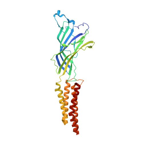
    GLIC is a bacterial homologue of the pentameric ligand-gated ion channels (pLGICs) that mediate the fast chemical neurotransmission of nerve signalling in eukaryotes. Because the activation and allosteric modulation features are conserved among prokaryotic and eukaryotic pLGICs, GLIC is commonly used as a model to study the allosteric transition and structural pharmacology of pLGICs. It has previously been shown that GLIC is inhibited by some carboxylic acid derivatives. Here, experimental evidence for carboxylate binding to GLIC is provided by solving its X-ray structures with a series of monocarboxylate and dicarboxylate derivatives, and two carboxylate-binding sites are described: (i) the `intersubunit' site that partially overlaps the canonical pLGIC orthosteric site and (ii) the `intrasubunit' vestibular site, which is only occupied by a subset of the described derivatives. While the intersubunit site is widely conserved in all pLGICs, the intrasubunit site is only conserved in cationic eukaryotic pLGICs. This study sheds light on the importance of these two extracellular modulation sites as potential drug targets in pLGICs.


  • Organizational Affiliation

    Unité Dynamique Structurale des Macromolécules, Institut Pasteur, 25 Rue du Docteur Roux, F-75015 Paris, France.


Macromolecules
Find similar proteins by:  (by identity cutoff)  |  3D Structure
Entity ID: 1
MoleculeChains Sequence LengthOrganismDetailsImage
Proton-gated ion channel
A, B, C, D, E
317Gloeobacter violaceus PCC 7421Mutation(s): 0 
Gene Names: glvIglr4197
Membrane Entity: Yes 
UniProt
Find proteins for Q7NDN8 (Gloeobacter violaceus (strain ATCC 29082 / PCC 7421))
Explore Q7NDN8 
Go to UniProtKB:  Q7NDN8
Entity Groups  
Sequence Clusters30% Identity50% Identity70% Identity90% Identity95% Identity100% Identity
UniProt GroupQ7NDN8
Sequence Annotations
Expand
  • Reference Sequence
Small Molecules
Ligands 4 Unique
IDChains Name / Formula / InChI Key2D Diagram3D Interactions
LMT
Query on LMT

Download Ideal Coordinates CCD File 
AA [auth D]
GA [auth E]
K [auth A]
L [auth A]
R [auth B]
AA [auth D],
GA [auth E],
K [auth A],
L [auth A],
R [auth B],
W [auth C]
DODECYL-BETA-D-MALTOSIDE
C24 H46 O11
NLEBIOOXCVAHBD-QKMCSOCLSA-N
PPI
Query on PPI

Download Ideal Coordinates CCD File 
BA [auth D]
CA [auth D]
DA [auth D]
HA [auth E]
M [auth A]
BA [auth D],
CA [auth D],
DA [auth D],
HA [auth E],
M [auth A],
N [auth A],
O [auth A],
S [auth B],
T [auth B],
X [auth C]
PROPANOIC ACID
C3 H6 O2
XBDQKXXYIPTUBI-UHFFFAOYSA-N
CL
Query on CL

Download Ideal Coordinates CCD File 
EA [auth E]
F [auth A]
G [auth A]
H [auth A]
P [auth B]
EA [auth E],
F [auth A],
G [auth A],
H [auth A],
P [auth B],
U [auth C],
Y [auth D]
CHLORIDE ION
Cl
VEXZGXHMUGYJMC-UHFFFAOYSA-M
NA
Query on NA

Download Ideal Coordinates CCD File 
FA [auth E]
I [auth A]
J [auth A]
Q [auth B]
V [auth C]
FA [auth E],
I [auth A],
J [auth A],
Q [auth B],
V [auth C],
Z [auth D]
SODIUM ION
Na
FKNQFGJONOIPTF-UHFFFAOYSA-N
Experimental Data & Validation

Experimental Data

  • Method: X-RAY DIFFRACTION
  • Resolution: 3.20 Å
  • R-Value Free: 0.201 
  • R-Value Work: 0.194 
  • R-Value Observed: 0.195 
  • Space Group: C 1 2 1
Unit Cell:
Length ( Å )Angle ( ˚ )
a = 180.69α = 90
b = 133.73β = 101.38
c = 158.89γ = 90
Software Package:
Software NamePurpose
BUSTERrefinement
XDSdata reduction
Aimlessdata scaling
REFMACphasing

Structure Validation

View Full Validation Report


Ligand Structure Quality Assessment 


Entry History & Funding Information

Deposition Data


Funding OrganizationLocationGrant Number
French National Research AgencyFrance13-BSV8-0020

Revision History  (Full details and data files)

  • Version 1.0: 2019-10-09
    Type: Initial release
  • Version 1.1: 2020-07-15
    Changes: Database references