


Crystal Structure of Arabidopsis thaliana UGT89C1 complexed with UDP
Zong, G.N., Wang, X.Q.To be published.
Experimental Data Snapshot
Entity ID: 1 | |||||
|---|---|---|---|---|---|
| Molecule | Chains | Sequence Length | Organism | Details | Image |
| UDP-glycosyltransferase 89C1 | 435 | Arabidopsis thaliana | Mutation(s): 0 EC: 2.4.1 | 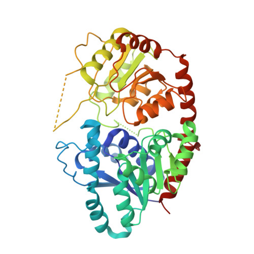 | |
UniProt | |||||
Find proteins for Q9LNE6 (Arabidopsis thaliana) Explore Q9LNE6 Go to UniProtKB: Q9LNE6 | |||||
Entity Groups | |||||
| Sequence Clusters | 30% Identity50% Identity70% Identity90% Identity95% Identity100% Identity | ||||
| UniProt Group | Q9LNE6 | ||||
Sequence AnnotationsExpand | |||||
| |||||
| Ligands 1 Unique | |||||
|---|---|---|---|---|---|
| ID | Chains | Name / Formula / InChI Key | 2D Diagram | 3D Interactions | |
| UDP Query on UDP | E [auth A], F [auth B], G [auth C], H [auth D] | URIDINE-5'-DIPHOSPHATE C9 H14 N2 O12 P2 XCCTYIAWTASOJW-XVFCMESISA-N |  | ||
| Length ( Å ) | Angle ( ˚ ) |
|---|---|
| a = 92.979 | α = 90 |
| b = 84.141 | β = 108.9 |
| c = 129.146 | γ = 90 |
| Software Name | Purpose |
|---|---|
| SCALEPACK | data scaling |
| REFMAC | refinement |
| PDB_EXTRACT | data extraction |
| HKL-2000 | data reduction |
| PHENIX | phasing |
| Funding Organization | Location | Grant Number |
|---|---|---|
| National Natural Science Foundation of China | China | 2017YFA0504801 |
RCSB PDB (citation) is hosted by
RCSB PDB is a member of the

