6L3H

Cryo-EM structure of dimeric quinol dependent Nitric Oxide Reductase (qNOR) from the pathogen Neisseria meninigitidis


Experimental Data Snapshot

  • Method: ELECTRON MICROSCOPY
  • Resolution: 3.06 Å
  • Aggregation State: PARTICLE 
  • Reconstruction Method: SINGLE PARTICLE 

wwPDB Validation   3D Report Full Report


This is version 1.2 of the entry. See complete history


Literature

The active form of quinol-dependent nitric oxide reductase fromNeisseria meningitidisis a dimer.

Jamali, M.A.M.Gopalasingam, C.C.Johnson, R.M.Tosha, T.Muramoto, K.Muench, S.P.Antonyuk, S.V.Shiro, Y.Hasnain, S.S.

(2020) IUCrJ 7: 404-415

  • DOI: https://doi.org/10.1107/S2052252520003656
  • Primary Citation of Related Structures:  
    6L1X, 6L3H, 6T6V

  • PubMed Abstract: 

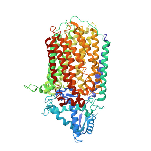
    Neisseria meningitidis is carried by nearly a billion humans, causing developmental impairment and over 100 000 deaths a year. A quinol-dependent nitric oxide reductase (qNOR) plays a critical role in the survival of the bacterium in the human host. X-ray crystallographic analyses of qNOR, including that from N. meningitidis ( Nm qNOR) reported here at 3.15 Å resolution, show monomeric assemblies, despite the more active dimeric sample being used for crystallization. Cryo-electron microscopic analysis of the same chromatographic fraction of Nm qNOR, however, revealed a dimeric assembly at 3.06 Å resolution. It is shown that zinc (which is used in crystallization) binding near the dimer-stabilizing TMII region contributes to the disruption of the dimer. A similar destabilization is observed in the monomeric (∼85 kDa) cryo-EM structure of a mutant (Glu494Ala) qNOR from the opportunistic pathogen Alcaligenes ( Achromobacter ) xylosoxidans , which primarily migrates as a monomer. The monomer-dimer transition of qNORs seen in the cryo-EM and crystallographic structures has wider implications for structural studies of multimeric membrane proteins. X-ray crystallographic and cryo-EM structural analyses have been performed on the same chromatographic fraction of Nm qNOR to high resolution. This represents one of the first examples in which the two approaches have been used to reveal a monomeric assembly in crystallo and a dimeric assembly in vitrified cryo-EM grids. A number of factors have been identified that may trigger the destabilization of helices that are necessary to preserve the integrity of the dimer. These include zinc binding near the entry of the putative proton-transfer channel and the preservation of the conformational integrity of the active site. The mutation near the active site results in disruption of the active site, causing an additional destabilization of helices (TMIX and TMX) that flank the proton-transfer channel helices, creating an inert monomeric enzyme.


  • Organizational Affiliation

    Graduate School of Life Science, University of Hyogo, 3-2-1 Kouto, Kamigori, Ako, Hyogo 678-1297, Japan.


Macromolecules
Find similar proteins by:  (by identity cutoff)  |  3D Structure
Entity ID: 1
MoleculeChains Sequence LengthOrganismDetailsImage
Nitric-oxide reductase
A, B
751Neisseria meningitidis alpha14Mutation(s): 0 
Gene Names: norBNMO_1451
EC: 1.7.99.7
Membrane Entity: Yes 
UniProt
Find proteins for C6S880 (Neisseria meningitidis (strain alpha14))
Explore C6S880 
Go to UniProtKB:  C6S880
Entity Groups  
Sequence Clusters30% Identity50% Identity70% Identity90% Identity95% Identity100% Identity
UniProt GroupC6S880
Sequence Annotations
Expand
  • Reference Sequence
Small Molecules
Ligands 3 Unique
IDChains Name / Formula / InChI Key2D Diagram3D Interactions
HEM
Query on HEM

Download Ideal Coordinates CCD File 
C [auth A],
D [auth A],
G [auth B],
H [auth B]
PROTOPORPHYRIN IX CONTAINING FE
C34 H32 Fe N4 O4
KABFMIBPWCXCRK-RGGAHWMASA-L
FE
Query on FE

Download Ideal Coordinates CCD File 
E [auth A],
I [auth B]
FE (III) ION
Fe
VTLYFUHAOXGGBS-UHFFFAOYSA-N
CA
Query on CA

Download Ideal Coordinates CCD File 
F [auth A],
J [auth B]
CALCIUM ION
Ca
BHPQYMZQTOCNFJ-UHFFFAOYSA-N
Experimental Data & Validation

Experimental Data

  • Method: ELECTRON MICROSCOPY
  • Resolution: 3.06 Å
  • Aggregation State: PARTICLE 
  • Reconstruction Method: SINGLE PARTICLE 
EM Software:
TaskSoftware PackageVersion
RECONSTRUCTIONRELION3.0
MODEL REFINEMENTPHENIX1.1.15

Structure Validation

View Full Validation Report


Entry History & Funding Information

Deposition Data


Funding OrganizationLocationGrant Number
Japan Society for the Promotion of Science (JSPS)JapanJP15K07029
Japan Society for the Promotion of Science (JSPS)JapanJP18K06162
Biotechnology and Biological Sciences Research Council (BBSRC)United KingdomBB/L006960/1
Biotechnology and Biological Sciences Research Council (BBSRC)United KingdomBB/N013972/1
Wellcome TrustUnited Kingdom108466/Z/15/Z
Wellcome TrustUnited Kingdom109158/B/15/Z

Revision History  (Full details and data files)

  • Version 1.0: 2020-04-01
    Type: Initial release
  • Version 1.1: 2020-06-03
    Changes: Database references
  • Version 1.2: 2024-03-27
    Changes: Data collection, Database references, Derived calculations, Refinement description