6YEO

Crystal structure of AmpC from E. coli with cyclic boronate 2


Experimental Data Snapshot

  • Method: X-RAY DIFFRACTION
  • Resolution: 2.04 Å
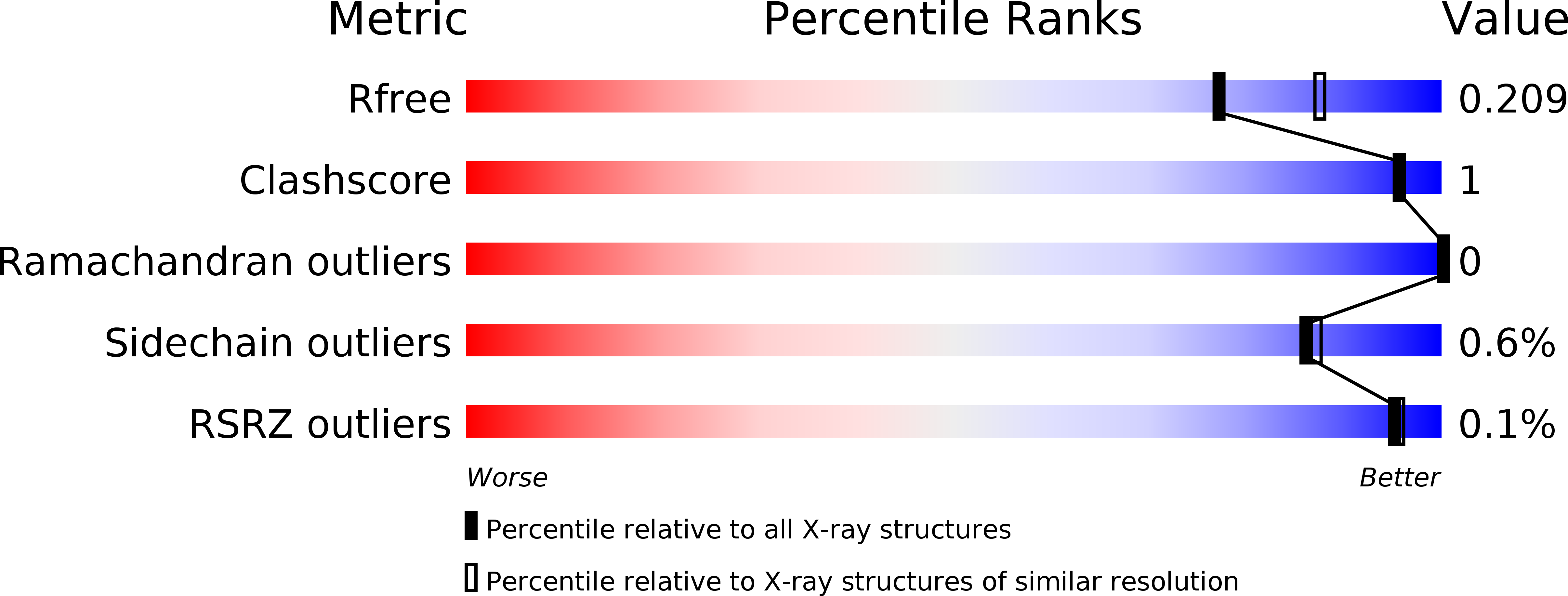
  • R-Value Free: 0.203 
  • R-Value Work: 0.172 
  • R-Value Observed: 0.173 

wwPDB Validation   3D Report Full Report


Ligand Structure Quality Assessment 


This is version 1.3 of the entry. See complete history


Literature

Bicyclic Boronates as Potent Inhibitors of AmpC, the Class C beta-Lactamase from Escherichia coli .

Lang, P.A.Parkova, A.Leissing, T.M.Calvopina, K.Cain, R.Krajnc, A.Panduwawala, T.D.Philippe, J.Fishwick, C.W.G.Trapencieris, P.Page, M.G.P.Schofield, C.J.Brem, J.

(2020) Biomolecules 10

  • DOI: https://doi.org/10.3390/biom10060899
  • Primary Citation of Related Structures:  
    6T3D, 6YEN, 6YEO, 6YPD

  • PubMed Abstract: 

    Resistance to β-lactam antibacterials, importantly via production of β-lactamases, threatens their widespread use. Bicyclic boronates show promise as clinically useful, dual-action inhibitors of both serine- (SBL) and metallo- (MBL) β-lactamases. In combination with cefepime, the bicyclic boronate taniborbactam is in phase 3 clinical trials for treatment of complicated urinary tract infections. We report kinetic and crystallographic studies on the inhibition of AmpC, the class C β‑lactamase from Escherichia coli , by bicyclic boronates, including taniborbactam, with different C-3 side chains. The combined studies reveal that an acylamino side chain is not essential for potent AmpC inhibition by active site binding bicyclic boronates. The tricyclic form of taniborbactam was observed bound to the surface of crystalline AmpC, but not at the active site, where the bicyclic form was observed. Structural comparisons reveal insights into why active site binding of a tricyclic form has been observed with the NDM-1 MBL, but not with other studied β-lactamases. Together with reported studies on the structural basis of inhibition of class A, B and D β‑lactamases, our data support the proposal that bicyclic boronates are broad-spectrum β‑lactamase inhibitors that work by mimicking a high energy 'tetrahedral' intermediate. These results suggest further SAR guided development could improve the breadth of clinically useful β-lactamase inhibition.


  • Organizational Affiliation

    Department of Chemistry, University of Oxford, Chemistry Research Laboratory, Oxford OX1 3TA, UK.


Macromolecules
Find similar proteins by:  (by identity cutoff)  |  3D Structure
Entity ID: 1
MoleculeChains Sequence LengthOrganismDetailsImage
Beta-lactamase
A, B, C, D
358Escherichia coli K-12Mutation(s): 0 
Gene Names: ampCampAb4150JW4111
EC: 3.5.2.6
UniProt
Find proteins for P00811 (Escherichia coli (strain K12))
Explore P00811 
Go to UniProtKB:  P00811
Entity Groups  
Sequence Clusters30% Identity50% Identity70% Identity90% Identity95% Identity100% Identity
UniProt GroupP00811
Sequence Annotations
Expand
  • Reference Sequence
Small Molecules
Ligands 6 Unique
IDChains Name / Formula / InChI Key2D Diagram3D Interactions
OK3 (Subject of Investigation/LOI)
Query on OK3

Download Ideal Coordinates CCD File 
E [auth A],
J [auth B],
N [auth C],
R [auth D]
(4~{R})-4-[[4-(aminomethyl)phenyl]carbonylamino]-3,3-bis(oxidanyl)-2-oxa-3-boranuidabicyclo[4.4.0]deca-1(10),6,8-triene-10-carboxylic acid
C17 H18 B N2 O6
QLXKUYDHLJOWQG-AWEZNQCLSA-N
EPE
Query on EPE

Download Ideal Coordinates CCD File 
G [auth A],
T [auth D]
4-(2-HYDROXYETHYL)-1-PIPERAZINE ETHANESULFONIC ACID
C8 H18 N2 O4 S
JKMHFZQWWAIEOD-UHFFFAOYSA-N
MPD
Query on MPD

Download Ideal Coordinates CCD File 
F [auth A],
K [auth B],
S [auth D]
(4S)-2-METHYL-2,4-PENTANEDIOL
C6 H14 O2
SVTBMSDMJJWYQN-YFKPBYRVSA-N
GOL
Query on GOL

Download Ideal Coordinates CCD File 
L [auth B],
O [auth C],
P [auth C],
Q [auth C]
GLYCEROL
C3 H8 O3
PEDCQBHIVMGVHV-UHFFFAOYSA-N
CL
Query on CL

Download Ideal Coordinates CCD File 
I [auth A],
M [auth B],
U [auth D]
CHLORIDE ION
Cl
VEXZGXHMUGYJMC-UHFFFAOYSA-M
NA
Query on NA

Download Ideal Coordinates CCD File 
H [auth A]SODIUM ION
Na
FKNQFGJONOIPTF-UHFFFAOYSA-N
Experimental Data & Validation

Experimental Data

  • Method: X-RAY DIFFRACTION
  • Resolution: 2.04 Å
  • R-Value Free: 0.203 
  • R-Value Work: 0.172 
  • R-Value Observed: 0.173 
  • Space Group: P 2 2 21
Unit Cell:
Length ( Å )Angle ( ˚ )
a = 100.683α = 90
b = 179.59β = 90
c = 99.658γ = 90
Software Package:
Software NamePurpose
XDSdata reduction
Aimlessdata scaling
PHENIXrefinement
PDB_EXTRACTdata extraction
PHASERphasing

Structure Validation

View Full Validation Report


Ligand Structure Quality Assessment 


Entry History & Funding Information

Deposition Data


Funding OrganizationLocationGrant Number
Medical Research Council (MRC, United Kingdom)United KingdomMRF-145-0004-TPG-AVISO

Revision History  (Full details and data files)

  • Version 1.0: 2020-06-24
    Type: Initial release
  • Version 1.1: 2020-07-01
    Changes: Database references
  • Version 1.2: 2020-09-30
    Changes: Database references
  • Version 1.3: 2024-01-24
    Changes: Data collection, Database references, Refinement description