pH-Induced Conformational Changes of Human Bocavirus Capsids.
Luo, M., Mietzsch, M., Chipman, P., Song, K., Xu, C., Spear, J., Sousa, D., McKenna, R., Soderlund-Venermo, M., Agbandje-McKenna, M.(2021) J Virol 95
- PubMed: 33472934
- DOI: https://doi.org/10.1128/JVI.02329-20
- Primary Citation of Related Structures:
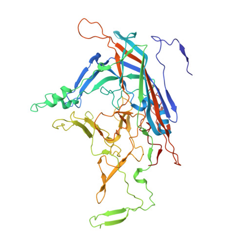
7L0U, 7L0V, 7L0W, 7L0X, 7L0Y - PubMed Abstract:
Human bocavirus 1 (HBoV1) and HBoV2-4 infect children and immunocompromised individuals, resulting in respiratory and gastrointestinal infections, respectively. Using cryo-electron microscopy and image reconstruction, the HBoV2 capsid structure was determined to 2.7 Å resolution at pH 7.4 and compared to the previously determined HBoV1, HBoV3, and HBoV4 structures. Consistent with previous findings, surface variable region (VR) III of the capsid protein VP3, proposed as a host tissue-tropism determinant, was structurally similar among the gastrointestinal strains HBoV2-4, but differed from HBoV1 with its tropism for the respiratory tract. Towards understanding the entry and trafficking properties of these viruses, HBoV1 and HBoV2 were further analyzed as species representatives of the two HBoV tropisms. Their cell surface glycan-binding characteristics were analyzed, and capsid structures determined to 2.5-2.7 Å resolution at pH 5.5 and 2.6, conditions normally encountered during infection. The data showed that glycans with terminal sialic acid, galactose, GlcNAc or heparan sulfate moieties do not facilitate HBoV1 or HBoV2 cellular attachment. With respect to trafficking, conformational changes common to both viruses were observed at low pH conditions localized to the VP N-terminus under the 5-fold channel, in the surface loops VR-I and VR-V and specific side-chain residues such as cysteines and histidines. The 5-fold conformational movements provide insight into the potential mechanism of VP N-terminal dynamics during HBoV infection and side-chain modifications highlight pH-sensitive regions of the capsid. IMPORTANCE Human bocaviruses (HBoVs) are associated with disease in humans. However, the lack of an animal model and a versatile cell culture system to study their life cycle limits the ability to develop specific treatments or vaccines. This study presents the structure of HBoV2, at 2.7 Å resolution, determined for comparison to the existing HBoV1, HBoV3, and HBoV4 structures, to enable the molecular characterization of strain and genus-specific capsid features contributing to tissue tropism and antigenicity. Furthermore, HBoV1 and HBoV2 structures determined under acidic conditions provide insight into capsid changes associated with endosomal and gastrointestinal acidification. Structural rearrangements of the capsid VP N-terminus, at the base of the 5-fold channel, demonstrate a disordering of a "basket" motif as pH decreases. These observations begin to unravel the molecular mechanism of HBoV infection and provide information for control strategies.
Organizational Affiliation:
Department of Biochemistry and Molecular Biology, Center for Structural Biology, McKnight Brain Institute, College of Medicine, Gainesville, FL 32610.