7NDP

X-ray structure of acetylcholine-binding protein (AChBP) in complex with FL001856.


Experimental Data Snapshot

  • Method: X-RAY DIFFRACTION
  • Resolution: 2.00 Å
  • R-Value Free: 0.243 
  • R-Value Work: 0.212 
  • R-Value Observed: 0.214 

wwPDB Validation   3D Report Full Report


Ligand Structure Quality Assessment 


This is version 1.2 of the entry. See complete history


Literature

Discovery of fragments inducing conformational effects in dynamic proteins using a second-harmonic generation biosensor.

FitzGerald, E.A.Butko, M.T.Boronat, P.Cederfelt, D.Abramsson, M.Ludviksdottir, H.van Muijlwijk-Koezen, J.E.de Esch, I.J.P.Dobritzsch, D.Young, T.Danielson, U.H.

(2021) RSC Adv 11: 7527-7537

  • DOI: https://doi.org/10.1039/d0ra09844b
  • Primary Citation of Related Structures:  
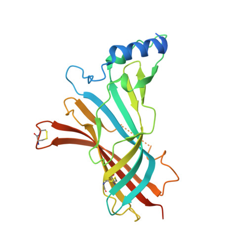
    7NDP

  • PubMed Abstract: 

    Biophysical screening of compound libraries for the identification of ligands that interact with a protein is efficient, but does typically not reveal if (or how) ligands may interfere with its functional properties. For this a biochemical/functional assay is required. But for proteins whose function is dependent on a conformational change, such assays are typically complex or have low throughput. Here we have explored a high-throughput second-harmonic generation (SHG) biosensor to detect fragments that induce conformational changes upon binding to a protein in real time and identify dynamic regions. Multiwell plate format SHG assays were developed for wild-type and six engineered single-cysteine mutants of acetyl choline binding protein (AChBP), a homologue to ligand gated ion channels (LGICs). They were conjugated with second harmonic-active labels via amine or maleimide coupling. To validate the assay, it was confirmed that the conformational changes induced in AChBP by nicotinic acetyl choline receptor (nAChR) agonists and antagonists were qualitatively different. A 1056 fragment library was subsequently screened against all variants and conformational modulators of AChBP were successfully identified, with hit rates from 9-22%, depending on the AChBP variant. A subset of four hits was selected for orthogonal validation and structural analysis. A time-resolved grating-coupled interferometry-based biosensor assay confirmed the interaction to be a reversible 1-step 1 : 1 interaction, and provided estimates of affinities and interaction kinetic rate constants ( K D = 0.28-63 μM, k a = 0.1-6 μM -1 s -1 , k d = 1 s -1 ). X-ray crystallography of two of the fragments confirmed their binding at a previously described conformationally dynamic site, corresponding to the regulatory site of LGICs. These results reveal that SHG has the sensitivity to identify fragments that induce conformational changes in a protein. A selection of fragment hits with a response profile different to known LGIC regulators was characterized and confirmed to bind to dynamic regions of the protein.


  • Organizational Affiliation

    Department of Chemistry - BMC, Uppsala University Uppsala 751 23 Sweden helena.danielson@kemi.uu.se.


Macromolecules
Find similar proteins by:  (by identity cutoff)  |  3D Structure
Entity ID: 1
MoleculeChains Sequence LengthOrganismDetailsImage
Acetylcholine-binding protein
A, B, C, D, E
A, B, C, D, E, F, G, H, I, J
237Lymnaea stagnalisMutation(s): 0 
UniProt
Find proteins for P58154 (Lymnaea stagnalis)
Explore P58154 
Go to UniProtKB:  P58154
Entity Groups  
Sequence Clusters30% Identity50% Identity70% Identity90% Identity95% Identity100% Identity
UniProt GroupP58154
Sequence Annotations
Expand
  • Reference Sequence
Oligosaccharides

Help

Entity ID: 2
MoleculeChains Length2D Diagram Glycosylation3D Interactions
2-acetamido-2-deoxy-beta-D-glucopyranose-(1-4)-2-acetamido-2-deoxy-beta-D-glucopyranose
K
2N-Glycosylation
Glycosylation Resources
GlyTouCan:  G42666HT
GlyCosmos:  G42666HT
GlyGen:  G42666HT
Small Molecules
Ligands 5 Unique
IDChains Name / Formula / InChI Key2D Diagram3D Interactions
U8T (Subject of Investigation/LOI)
Query on U8T

Download Ideal Coordinates CCD File 
BA [auth H]
CA [auth I]
FA [auth J]
P [auth B]
Q [auth C]
BA [auth H],
CA [auth I],
FA [auth J],
P [auth B],
Q [auth C],
T [auth D],
V [auth E],
Y [auth G]
6-bromanylspiro[3~{H}-chromene-2,4'-piperidine]-4-one
C13 H14 Br N O2
HTOKNOIQHUEHNB-UHFFFAOYSA-N
NAG
Query on NAG

Download Ideal Coordinates CCD File 
AA [auth H]
EA [auth J]
L [auth A]
O [auth B]
W [auth F]
AA [auth H],
EA [auth J],
L [auth A],
O [auth B],
W [auth F],
X [auth G]
2-acetamido-2-deoxy-beta-D-glucopyranose
C8 H15 N O6
OVRNDRQMDRJTHS-FMDGEEDCSA-N
SO4
Query on SO4

Download Ideal Coordinates CCD File 
Z [auth G]SULFATE ION
O4 S
QAOWNCQODCNURD-UHFFFAOYSA-L
GOL
Query on GOL

Download Ideal Coordinates CCD File 
M [auth A],
N [auth A],
R [auth D],
S [auth D],
U [auth E]
GLYCEROL
C3 H8 O3
PEDCQBHIVMGVHV-UHFFFAOYSA-N
CL
Query on CL

Download Ideal Coordinates CCD File 
DA [auth I],
GA [auth J]
CHLORIDE ION
Cl
VEXZGXHMUGYJMC-UHFFFAOYSA-M
Experimental Data & Validation

Experimental Data

  • Method: X-RAY DIFFRACTION
  • Resolution: 2.00 Å
  • R-Value Free: 0.243 
  • R-Value Work: 0.212 
  • R-Value Observed: 0.214 
  • Space Group: P 21 21 21
Unit Cell:
Length ( Å )Angle ( ˚ )
a = 76.7α = 90
b = 121.1β = 90
c = 241.3γ = 90
Software Package:
Software NamePurpose
REFMACrefinement
PHASERphasing
XSCALEdata scaling
XDSdata reduction

Structure Validation

View Full Validation Report


Ligand Structure Quality Assessment 


Entry History & Funding Information

Deposition Data


Funding OrganizationLocationGrant Number
Marie Sklodowska-Curie Actions, FragNET ITNEuropean Union675899

Revision History  (Full details and data files)

  • Version 1.0: 2021-04-07
    Type: Initial release
  • Version 1.1: 2022-04-27
    Changes: Database references
  • Version 1.2: 2024-01-31
    Changes: Data collection, Refinement description