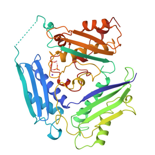
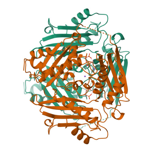
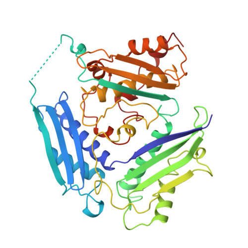
1XRC

CRYSTAL STRUCTURE OF S-ADENOSYLMETHIONINE SYNTHETASE


Experimental Data Snapshot

  • Method: X-RAY DIFFRACTION
  • Resolution: 3.00 Å
  • R-Value Free: 0.258 
  • R-Value Work: 0.197 
  • R-Value Observed: 0.197 

wwPDB Validation   3D Report Full Report


This is version 1.3 of the entry. See complete history


Literature

Crystal structure of S-adenosylmethionine synthetase.

Takusagawa, F.Kamitori, S.Misaki, S.Markham, G.D.

(1996) J Biol Chem 271: 136-147

  • Primary Citation of Related Structures:  
    1XRA, 1XRB, 1XRC

  • PubMed Abstract: 

    The structure of S-adenosylmethionine synthetase (MAT, ATP:L-methionine S-adenosyltransferase, EC 2.5.1.6.) from Escherichia coli has been determined at 3.0 A resolution by multiple isomorphous replacement using a uranium derivative and the selenomethionine form of the enzyme (SeMAT). The SeMAT data (9 selenomethionine residues out of 383 amino acid residues) have been found to have a sufficient phasing power to determine the structure of the 42,000 molecular weight protein by combining them with the other heavy atom derivative data (multiple isomorphous replacement). The enzyme consists of four identical subunits; two subunits form a spherical tight dimer, and pairs of these dimers form a peanut-shaped tetrameric enzyme. Each pair dimer has two active sites which are located between the subunits. Each subunit consists of three domains that are related to each other by pseudo-3-fold symmetry. The essential divalent (Mg2+/Co2+) and monovalent (K+) metal ions and one of the product, Pi ions, were found in the active site from three separate structures.


  • Organizational Affiliation

    Department of Chemistry, University of Kansas, Lawrence 66045-0046, USA.


Macromolecules
Find similar proteins by:  (by identity cutoff)  |  3D Structure
Entity ID: 1
MoleculeChains Sequence LengthOrganismDetailsImage
S-ADENOSYLMETHIONINE SYNTHETASE383Escherichia coliMutation(s): 0 
EC: 2.5.1.6
UniProt
Find proteins for P0A817 (Escherichia coli (strain K12))
Explore P0A817 
Go to UniProtKB:  P0A817
Entity Groups  
Sequence Clusters30% Identity50% Identity70% Identity90% Identity95% Identity100% Identity
UniProt GroupP0A817
Sequence Annotations
Expand
  • Reference Sequence
Experimental Data & Validation

Experimental Data

  • Method: X-RAY DIFFRACTION
  • Resolution: 3.00 Å
  • R-Value Free: 0.258 
  • R-Value Work: 0.197 
  • R-Value Observed: 0.197 
  • Space Group: P 62 2 2
Unit Cell:
Length ( Å )Angle ( ˚ )
a = 128.9α = 90
b = 128.9β = 90
c = 139.8γ = 120
Software Package:
Software NamePurpose
X-PLORmodel building
X-PLORrefinement
X-PLORphasing

Structure Validation

View Full Validation Report


Entry History 

Deposition Data

Revision History  (Full details and data files)

  • Version 1.0: 1996-03-08
    Type: Initial release
  • Version 1.1: 2008-03-24
    Changes: Version format compliance
  • Version 1.2: 2011-07-13
    Changes: Version format compliance
  • Version 1.3: 2024-02-14
    Changes: Data collection, Database references, Derived calculations, Other