227D

A CRYSTALLOGRAPHIC AND SPECTROSCOPIC STUDY OF THE COMPLEX BETWEEN D(CGCGAATTCGCG)2 AND 2,5-BIS(4-GUANYLPHENYL)FURAN, AN ANALOGUE OF BERENIL. STRUCTURAL ORIGINS OF ENHANCED DNA-BINDING AFFINITY


Experimental Data Snapshot

  • Method: X-RAY DIFFRACTION
  • Resolution: 2.20 Å
  • R-Value Work: 0.179 
  • R-Value Observed: 0.179 

wwPDB Validation   3D Report Full Report


Ligand Structure Quality Assessment 


This is version 1.3 of the entry. See complete history


Literature

A crystallographic and spectroscopic study of the complex between d(CGCGAATTCGCG)2 and 2,5-bis(4-guanylphenyl)furan, an analogue of berenil. Structural origins of enhanced DNA-binding affinity.

Laughton, C.A.Tanious, F.Nunn, C.M.Boykin, D.W.Wilson, W.D.Neidle, S.

(1996) Biochemistry 35: 5655-5661

  • DOI: https://doi.org/10.1021/bi952162r
  • Primary Citation of Related Structures:  
    227D

  • PubMed Abstract: 

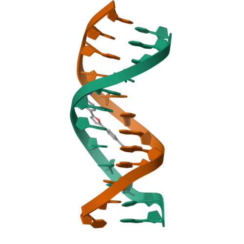
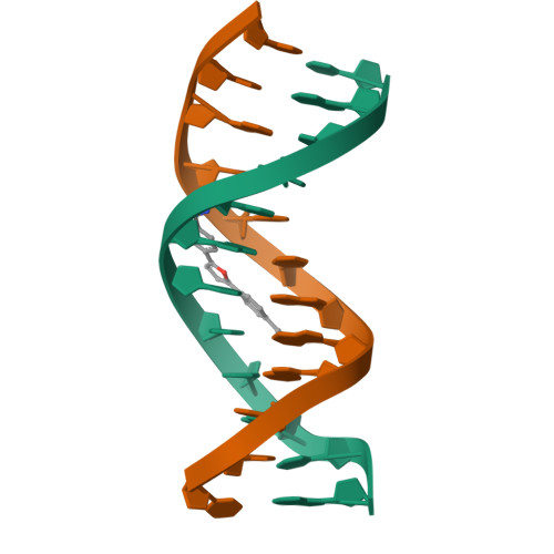
    2,5-Bis(4-guanylphenyl)furan ("furamidine") is a dicationic minor groove binding drug that has been shown to be more effective than pentamidine against the Pneumocystis carinii pathogen in an immunosuppressed rat model. It has a close structural similarity to the antitrypanosomal drug berenil, differing only on the replacement of the central triazene unit with a furan moiety. we have determined the crystal structure of the complex between furamidine and the DNA dodecamer d(CGCGAATTCGCG)2 and compared it with the same DNA sequence by UV-visible, fluorescence, and CD spectroscopy. Furamidine shows tighter binding to this sequence (Keq = 6.7 x 10(6)) than berenil (Keq = 6.6 x 10(5)). The crystal structure reveals that, unlike berenil, furamidine makes direct hydrogen bond interactions with this DNA sequence through both amidinium groups to O2 atoms of thymine bases and is more isohelical with the minor groove. Molecular mechanics calculations support the hypothesis that these differences result in the improved interaction energy between the ligand and the DNA.


  • Organizational Affiliation

    CRC Biomolecular Structure Unit, Institute of Cancer Research, Sutton Surrey, UK.


Macromolecules

Find similar nucleic acids by:  Sequence   |   3D Structure  

Entity ID: 1
MoleculeChains LengthOrganismImage
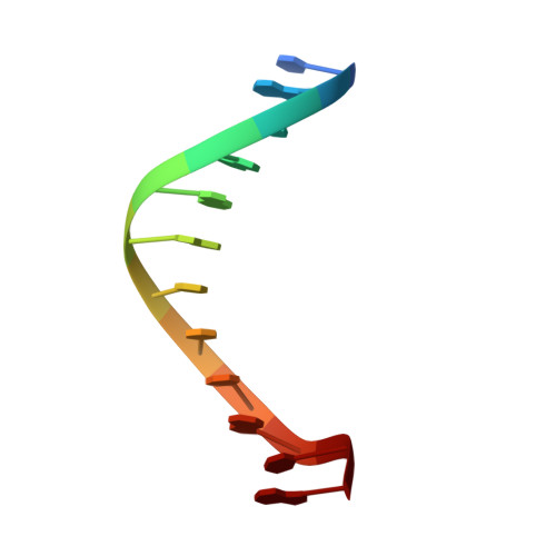
DNA (5'-D(*CP*GP*CP*GP*AP*AP*TP*TP*CP*GP*CP*G)-3')
A, B
12N/A
Sequence Annotations
Expand
  • Reference Sequence
Small Molecules
Ligands 1 Unique
IDChains Name / Formula / InChI Key2D Diagram3D Interactions
BGF
Query on BGF

Download Ideal Coordinates CCD File 
C [auth A]2,5-BIS(4-GUANYLPHENYL)FURAN
C18 H18 N4 O
ZJHZBDRZEZEDGB-UHFFFAOYSA-P
Experimental Data & Validation

Experimental Data

  • Method: X-RAY DIFFRACTION
  • Resolution: 2.20 Å
  • R-Value Work: 0.179 
  • R-Value Observed: 0.179 
  • Space Group: P 21 21 21
Unit Cell:
Length ( Å )Angle ( ˚ )
a = 25.28α = 90
b = 40.69β = 90
c = 66.73γ = 90
Software Package:
Software NamePurpose
X-PLORrefinement

Structure Validation

View Full Validation Report


Ligand Structure Quality Assessment 


Entry History 

Deposition Data

Revision History  (Full details and data files)

  • Version 1.0: 1995-11-11
    Type: Initial release
  • Version 1.1: 2008-05-22
    Changes: Version format compliance
  • Version 1.2: 2011-07-13
    Changes: Version format compliance
  • Version 1.3: 2024-02-14
    Changes: Data collection, Database references, Derived calculations