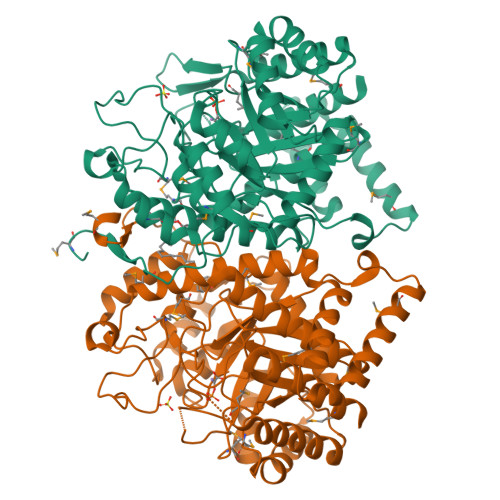
2B7O

The Structure of 3-Deoxy-D-Arabino-Heptulosonate 7-Phosphate Synthase from Mycobacterium tuberculosis


Experimental Data Snapshot

  • Method: X-RAY DIFFRACTION
  • Resolution: 2.30 Å
  • R-Value Free: 0.224 
  • R-Value Work: 0.188 
  • R-Value Observed: 0.190 

wwPDB Validation   3D Report Full Report


Ligand Structure Quality Assessment 


This is version 1.3 of the entry. See complete history


Literature

The structure of 3-deoxy-d-arabino-heptulosonate 7-phosphate synthase from Mycobacterium tuberculosis reveals a common catalytic scaffold and ancestry for type I and type II enzymes

Webby, C.J.Baker, H.M.Lott, J.S.Baker, E.N.Parker, E.J.

(2005) J Mol Biol 354: 927-939

  • DOI: https://doi.org/10.1016/j.jmb.2005.09.093
  • Primary Citation of Related Structures:  
    2B7O

  • PubMed Abstract: 

    The shikimate pathway, responsible for the biosynthesis of aromatic compounds, is essential for the growth of Mycobacterium tuberculosis and is a potential target for the design of new anti-tuberculosis drugs. The first step of this pathway is catalyzed by 3-deoxy-d-arabino-heptulosonate 7-phosphate synthase (DAH7PS). The DAH7PSs have been classified into two apparently unrelated types and, whereas structural data have been obtained for the type I DAH7PSs, no structural information is available for their type II counterparts. The type II DAH7PS from M.tuberculosis has been expressed in Escherichia coli, purified, functionally characterized and crystallized. It is found to be metal ion-dependent and subject to feedback inhibition by phenylalanine, tryptophan, tyrosine and chorismate, with a significant synergistic effect when tryptophan is used in combination with phenylalanine. The crystal structure of M.tuberculosis DAH7PS has been determined by single-wavelength anomalous diffraction and refined at 2.3A in complex with substrate phosphoenolpyruvate and Mn(2+). The structure reveals a tightly associated dimer of (beta/alpha)(8) TIM barrels. The monomer fold, the arrangement of key residues in the active site, and the binding modes of PEP and Mn(2+), all match those of the type I enzymes, and indicate a common ancestry for the type I and type II DAH7PSs, despite their minimal sequence identity. In contrast, the structural elements that decorate the core (beta/alpha)(8) fold differ from those in the type I enzymes, consistent with their different regulatory and oligomeric properties.


  • Organizational Affiliation

    Centre of Structural Biology, Institute of Fundamental Sciences, Massey University, Palmerston North, New Zealand.


Macromolecules
Find similar proteins by:  (by identity cutoff)  |  3D Structure
Entity ID: 1
MoleculeChains Sequence LengthOrganismDetailsImage
3-deoxy-D-arabino-heptulosonate 7-phosphate synthase AroG
A, B
464Mycobacterium tuberculosis H37RvMutation(s): 0 
Gene Names: Rv2178c
EC: 2.5.1.54
UniProt
Find proteins for O53512 (Mycobacterium tuberculosis (strain ATCC 25618 / H37Rv))
Go to UniProtKB:  O53512
Sequence Annotations
Expand
  • Reference Sequence
Small Molecules
Ligands 4 Unique
IDChains Name / Formula / InChI Key2D Diagram3D Interactions
CE1
Query on CE1

Download Ideal Coordinates CCD File 
F [auth A]O-DODECANYL OCTAETHYLENE GLYCOL
C28 H58 O9
YYELLDKEOUKVIQ-UHFFFAOYSA-N
PEP
Query on PEP

Download Ideal Coordinates CCD File 
E [auth A],
I [auth B]
PHOSPHOENOLPYRUVATE
C3 H5 O6 P
DTBNBXWJWCWCIK-UHFFFAOYSA-N
SO4
Query on SO4

Download Ideal Coordinates CCD File 
D [auth A],
G [auth B]
SULFATE ION
O4 S
QAOWNCQODCNURD-UHFFFAOYSA-L
MN
Query on MN

Download Ideal Coordinates CCD File 
C [auth A],
H [auth B]
MANGANESE (II) ION
Mn
WAEMQWOKJMHJLA-UHFFFAOYSA-N
Modified Residues  1 Unique
IDChains TypeFormula2D DiagramParent
MSE
Query on MSE
A, B
L-PEPTIDE LINKINGC5 H11 N O2 SeMET
Experimental Data & Validation

Experimental Data

  • Method: X-RAY DIFFRACTION
  • Resolution: 2.30 Å
  • R-Value Free: 0.224 
  • R-Value Work: 0.188 
  • R-Value Observed: 0.190 
  • Space Group: P 32 2 1
Unit Cell:
Length ( Å )Angle ( ˚ )
a = 204.085α = 90
b = 204.085β = 90
c = 66.23γ = 120
Software Package:
Software NamePurpose
REFMACrefinement
DENZOdata reduction
SCALEPACKdata scaling
SHELXSphasing

Structure Validation

View Full Validation Report


Ligand Structure Quality Assessment 


Entry History 

Revision History  (Full details and data files)

  • Version 1.0: 2005-10-18
    Type: Initial release
  • Version 1.1: 2008-04-25
    Changes: Version format compliance
  • Version 1.2: 2011-07-13
    Changes: Source and taxonomy, Version format compliance
  • Version 1.3: 2024-10-16
    Changes: Data collection, Database references, Derived calculations, Structure summary