Structure of N-acetyl-beta-D-glucosaminidase (GcnA) from the Endocarditis Pathogen Streptococcus gordonii and its Complex with the Mechanism-based Inhibitor NAG-thiazoline
Langley, D.B., Harty, D.W.S., Jacques, N.A., Hunter, N., Guss, J.M., Collyer, C.A.(2008) J Mol Biology 377: 104-116
- PubMed: 18237743
- DOI: https://doi.org/10.1016/j.jmb.2007.09.028
- Primary Citation of Related Structures:
2EPK, 2EPL, 2EPM, 2EPN, 2EPO - PubMed Abstract:
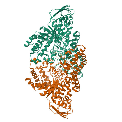
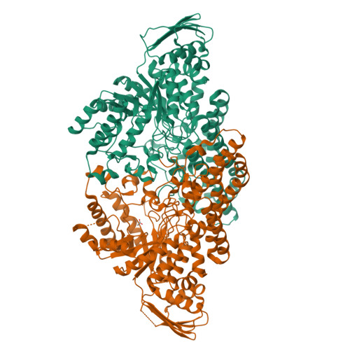
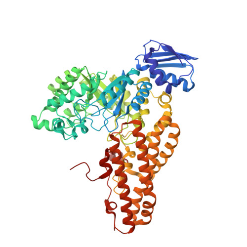
The crystal structure of GcnA, an N-acetyl-beta-D-glucosaminidase from Streptococcus gordonii, was solved by multiple wavelength anomalous dispersion phasing using crystals of selenomethionine-substituted protein. GcnA is a homodimer with subunits each comprised of three domains. The structure of the C-terminal alpha-helical domain has not been observed previously and forms a large dimerisation interface. The fold of the N-terminal domain is observed in all structurally related glycosidases although its function is unknown. The central domain has a canonical (beta/alpha)(8) TIM-barrel fold which harbours the active site. The primary sequence and structure of this central domain identifies the enzyme as a family 20 glycosidase. Key residues implicated in catalysis have different conformations in two different crystal forms, which probably represent active and inactive conformations of the enzyme. The catalytic mechanism for this class of glycoside hydrolase, where the substrate rather than the enzyme provides the cleavage-inducing nucleophile, has been confirmed by the structure of GcnA complexed with a putative reaction intermediate analogue, N-acetyl-beta-D-glucosamine-thiazoline. The catalytic mechanism is discussed in light of these and other family 20 structures.
Organizational Affiliation:
School of Molecular and Microbial Biosciences, University of Sydney, Sydney, Australia. d.langley@mmb.usyd.edu.au


