2FA1 | pdb_00002fa1

Crystal structure of PhnF C-terminal domain


Experimental Data Snapshot

  • Method: X-RAY DIFFRACTION
  • Resolution: 1.70 Å
  • R-Value Free: 
    0.241 (Depositor), 0.240 (DCC) 
  • R-Value Work: 
    0.187 (Depositor), 0.190 (DCC) 
  • R-Value Observed: 
    0.190 (Depositor) 

wwPDB Validation   3D Report Full Report


Ligand Structure Quality Assessment 

Created with Raphaël 2.3.0Worse 01 BetterLigand structure goodness of fit to experimental dataBest fitted BDFClick on this verticalbar to view details

This is version 1.5 of the entry. See complete history


Literature

Structural characterization of GntR/HutC family signaling domain.

Gorelik, M.Lunin, V.V.Skarina, T.Savchenko, A.

(2006) Protein Sci 15: 1-6

  • DOI: https://doi.org/10.1110/ps.062146906
  • Primary Citation of Related Structures:  
    2FA1

  • PubMed Abstract: 

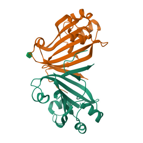
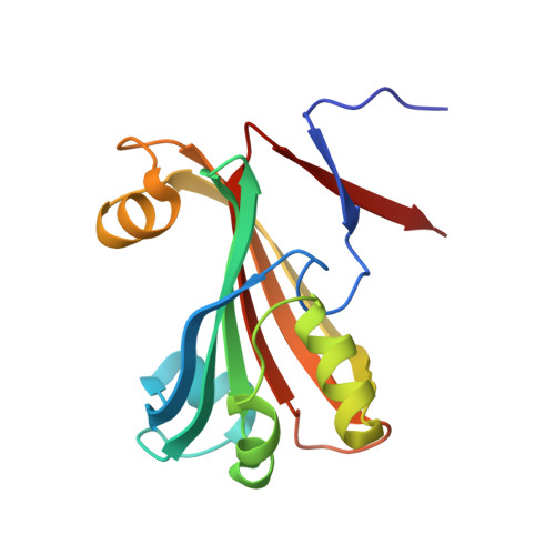
    The crystal structure of Escherichia coli PhnF C-terminal domain (C-PhnF) was solved at 1.7 A resolution by the single wavelength anomalous dispersion (SAD) method. The PhnF protein belongs to the HutC subfamily of the large GntR transcriptional regulator family. Members of this family share similar N-terminal DNA-binding domains, but are divided into four subfamilies according to their heterogenic C-terminal domains, which are involved in effector binding and oligomerization. The C-PhnF structure provides for the first time the scaffold of this domain for the HutC subfamily, which covers about 31% of GntR-like regulators. The structure represents a mixture of alpha-helices and beta-strands, with a six-stranded antiparallel beta-sheet at the core. C-PhnF monomers form a dimer by establishing interdomain eight-strand beta-sheets that include core antiparallel and N-terminal two-strand parallel beta-sheets from each monomer. C-PhnF shares strong structural similarity with the chorismate lyase fold, which features a buried active site locked behind two helix-turn-helix loops. The structural comparison of the C-PhnF and UbiC proteins allows us to propose that a similar site in the PhnF structure is adapted for effector binding.


  • Organizational Affiliation

    Ontario Center for Structural Proteomics, University Health Network, University of Toronto, Toronto, Ontario M5G 1L7, Canada.


Macromolecules
Find similar proteins by:  (by identity cutoff)  |  3D Structure
Entity ID: 1
MoleculeChains Sequence LengthOrganismDetailsImage
Probable transcriptional regulator phnF
A, B
160Escherichia coliMutation(s): 0 
Gene Names: phnF
UniProt
Find proteins for P16684 (Escherichia coli (strain K12))
Explore P16684 
Go to UniProtKB:  P16684
Entity Groups  
Sequence Clusters30% Identity50% Identity70% Identity90% Identity95% Identity100% Identity
UniProt GroupP16684
Sequence Annotations
Expand
  • Reference Sequence
Small Molecules
Ligands 1 Unique
IDChains Name / Formula / InChI Key2D Diagram3D Interactions
BDF
Query on BDF

Download Ideal Coordinates CCD File 
C [auth B]beta-D-fructopyranose
C6 H12 O6
LKDRXBCSQODPBY-ARQDHWQXSA-N
Experimental Data & Validation

Experimental Data

  • Method: X-RAY DIFFRACTION
  • Resolution: 1.70 Å
  • R-Value Free:  0.241 (Depositor), 0.240 (DCC) 
  • R-Value Work:  0.187 (Depositor), 0.190 (DCC) 
  • R-Value Observed: 0.190 (Depositor) 
Space Group: P 1 21 1
Unit Cell:
Length ( Å )Angle ( ˚ )
a = 46.765α = 90
b = 51.572β = 103.64
c = 63.961γ = 90
Software Package:
Software NamePurpose
REFMACrefinement
HKL-2000data reduction
SCALEPACKdata scaling
SOLVEphasing

Structure Validation

View Full Validation Report


Ligand Structure Quality Assessment 

Created with Raphaël 2.3.0Worse 01 BetterLigand structure goodness of fit to experimental dataBest fitted BDFClick on this verticalbar to view details

Entry History 

Revision History  (Full details and data files)

  • Version 1.0: 2006-01-10
    Type: Initial release
  • Version 1.1: 2008-05-01
    Changes: Version format compliance
  • Version 1.2: 2011-07-13
    Changes: Version format compliance
  • Version 1.3: 2011-10-05
    Changes: Structure summary
  • Version 1.4: 2020-07-29
    Type: Remediation
    Reason: Carbohydrate remediation
    Changes: Data collection, Database references, Derived calculations, Structure summary
  • Version 1.5: 2024-02-14
    Changes: Data collection, Database references, Structure summary