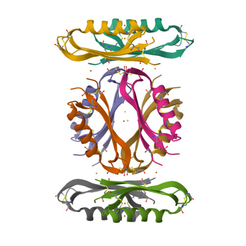
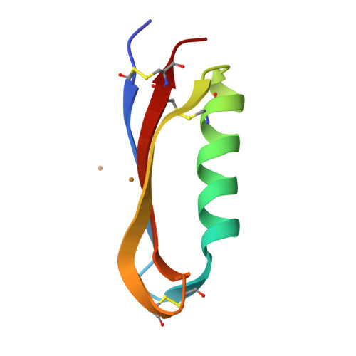
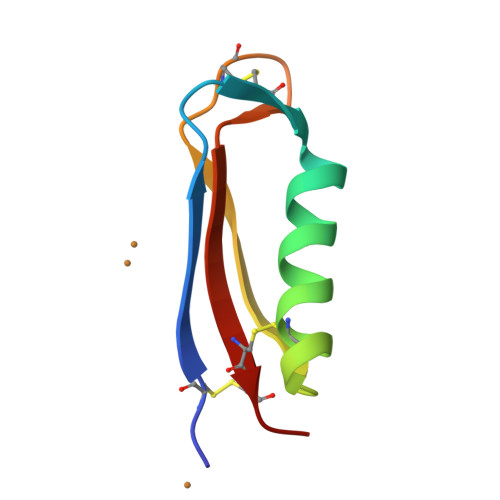
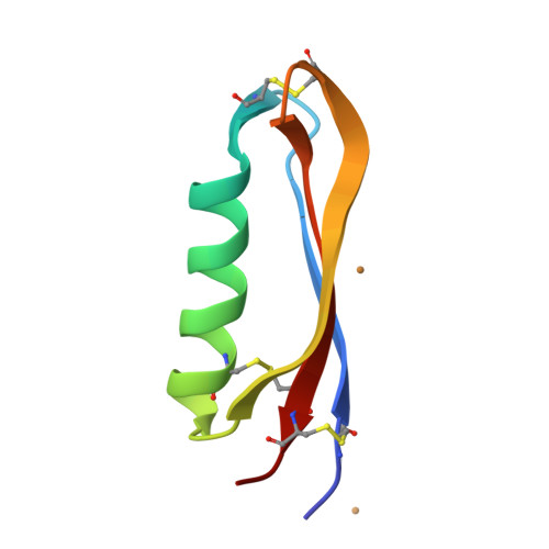
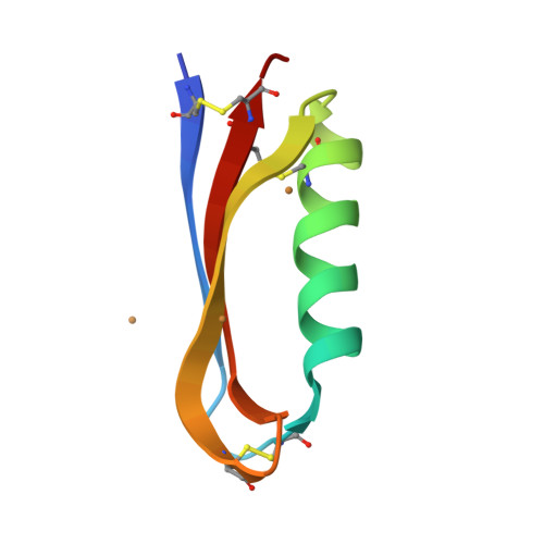
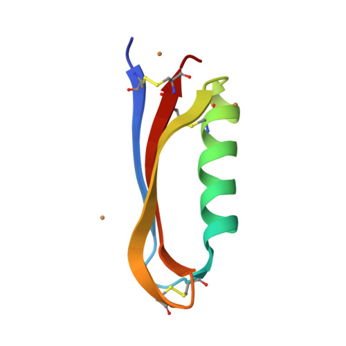
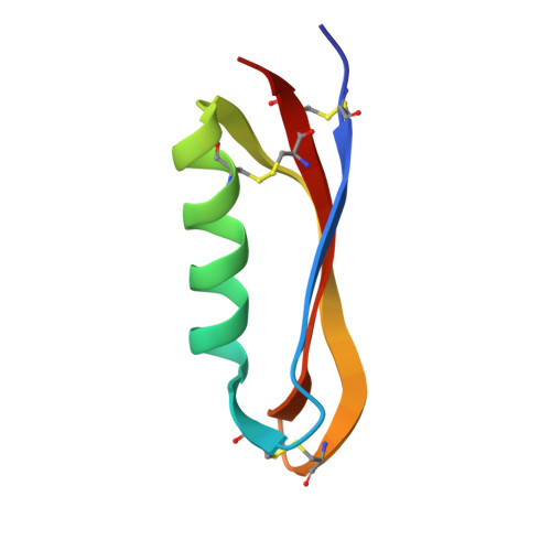
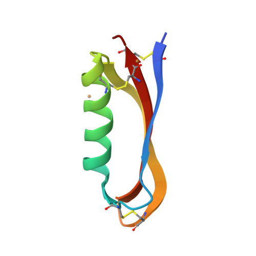
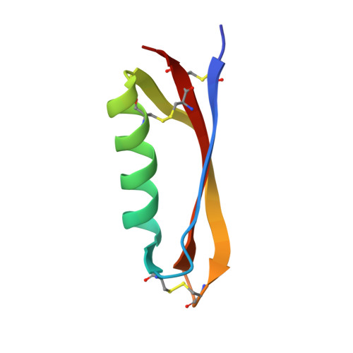
2FK3

Structure of the Alzheimer's Amyloid Precursor Protein (APP) Copper Binding Domain in 'large unit cell' form


Experimental Data Snapshot

  • Method: X-RAY DIFFRACTION
  • Resolution: 2.40 Å
  • R-Value Free: 0.248 
  • R-Value Work: 0.208 
  • R-Value Observed: 0.208 

wwPDB Validation   3D Report Full Report


This is version 1.4 of the entry. See complete history


Literature

Structural Studies of the Alzheimer's Amyloid Precursor Protein Copper-binding Domain Reveal How it Binds Copper Ions

Kong, G.K.Adams, J.J.Harris, H.H.Boas, J.F.Curtain, C.C.Galatis, D.Masters, C.L.Barnham, K.J.McKinstry, W.J.Cappai, R.Parker, M.W.

(2007) J Mol Biol 367: 148-161

  • DOI: https://doi.org/10.1016/j.jmb.2006.12.041
  • Primary Citation of Related Structures:  
    2FJZ, 2FK1, 2FK2, 2FK3, 2FKL

  • PubMed Abstract: 

    Alzheimer's disease (AD) is the major cause of dementia. Amyloid beta peptide (Abeta), generated by proteolytic cleavage of the amyloid precursor protein (APP), is central to AD pathogenesis. APP can function as a metalloprotein and modulate copper (Cu) transport, presumably via its extracellular Cu-binding domain (CuBD). Cu binding to the CuBD reduces Abeta levels, suggesting that a Cu mimetic may have therapeutic potential. We describe here the atomic structures of apo CuBD from three crystal forms and found they have identical Cu-binding sites despite the different crystal lattices. The structure of Cu(2+)-bound CuBD reveals that the metal ligands are His147, His151, Tyr168 and two water molecules, which are arranged in a square pyramidal geometry. The site resembles a Type 2 non-blue Cu center and is supported by electron paramagnetic resonance and extended X-ray absorption fine structure studies. A previous study suggested that Met170 might be a ligand but we suggest that this residue plays a critical role as an electron donor in CuBDs ability to reduce Cu ions. The structure of Cu(+)-bound CuBD is almost identical to the Cu(2+)-bound structure except for the loss of one of the water ligands. The geometry of the site is unfavorable for Cu(+), thus providing a mechanism by which CuBD could readily transfer Cu ions to other proteins.


  • Organizational Affiliation

    Biota Structural Biology Laboratory, St. Vincent's Institute, 9 Princes Street, Fitzroy, Victoria 3065, Australia.


Macromolecules
Find similar proteins by:  (by identity cutoff)  |  3D Structure
Entity ID: 1
MoleculeChains Sequence LengthOrganismDetailsImage
Amyloid beta A4 protein precursor
A, B, C, D, E
A, B, C, D, E, F, G, H
59Homo sapiensMutation(s): 0 
Gene Names: APP
UniProt & NIH Common Fund Data Resources
Find proteins for P05067 (Homo sapiens)
Explore P05067 
Go to UniProtKB:  P05067
PHAROS:  P05067
GTEx:  ENSG00000142192 
Entity Groups  
Sequence Clusters30% Identity50% Identity70% Identity90% Identity95% Identity100% Identity
UniProt GroupP05067
Sequence Annotations
Expand
  • Reference Sequence
Small Molecules
Ligands 1 Unique
IDChains Name / Formula / InChI Key2D Diagram3D Interactions
CU
Query on CU

Download Ideal Coordinates CCD File 
AA [auth H]
I [auth A]
J [auth A]
K [auth A]
L [auth A]
AA [auth H],
I [auth A],
J [auth A],
K [auth A],
L [auth A],
M [auth B],
N [auth B],
O [auth C],
P [auth C],
Q [auth C],
R [auth D],
S [auth D],
T [auth E],
U [auth E],
V [auth E],
W [auth F],
X [auth F],
Y [auth F],
Z [auth H]
COPPER (II) ION
Cu
JPVYNHNXODAKFH-UHFFFAOYSA-N
Experimental Data & Validation

Experimental Data

  • Method: X-RAY DIFFRACTION
  • Resolution: 2.40 Å
  • R-Value Free: 0.248 
  • R-Value Work: 0.208 
  • R-Value Observed: 0.208 
  • Space Group: P 21 21 21
Unit Cell:
Length ( Å )Angle ( ˚ )
a = 59.623α = 90
b = 67.437β = 90
c = 127.656γ = 90
Software Package:
Software NamePurpose
CNSrefinement
DENZOdata reduction
SCALEPACKdata scaling
SOLVEphasing

Structure Validation

View Full Validation Report


Entry History 

Deposition Data

Revision History  (Full details and data files)

  • Version 1.0: 2007-01-16
    Type: Initial release
  • Version 1.1: 2008-05-01
    Changes: Version format compliance
  • Version 1.2: 2011-07-13
    Changes: Version format compliance
  • Version 1.3: 2022-02-23
    Changes: Data collection, Database references, Derived calculations
  • Version 1.4: 2024-11-13
    Changes: Data collection, Structure summary