2PMV

Crystal Structure of Human Intrinsic Factor- Cobalamin Complex at 2.6 A Resolution


Experimental Data Snapshot

  • Method: X-RAY DIFFRACTION
  • Resolution: 2.60 Å
  • R-Value Free: 0.249 
  • R-Value Work: 0.213 
  • R-Value Observed: 0.213 

wwPDB Validation   3D Report Full Report


Ligand Structure Quality Assessment 


This is version 2.1 of the entry. See complete history


Literature

Crystal structure of human intrinsic factor: Cobalamin complex at 2.6-A resolution

Mathews, F.S.Gordon, M.M.Chen, Z.Rajashankar, K.R.Ealick, S.E.Alpers, D.H.Sukumar, N.

(2007) Proc Natl Acad Sci U S A 104: 17311-17316

  • DOI: https://doi.org/10.1073/pnas.0703228104
  • Primary Citation of Related Structures:  
    2PMV

  • PubMed Abstract: 

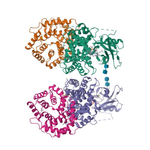
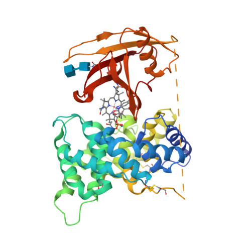
    The structure of intrinsic factor (IF) in complex with cobalamin (Cbl) was determined at 2.6-A resolution. The overall fold of the molecule is that of an alpha(6)/alpha(6) barrel. It is a two-domain protein, and the Cbl is bound at the interface of the domains in a base-on conformation. Surprisingly, two full-length molecules, each comprising an alpha- and a beta-domain and one Cbl, and two truncated molecules with only an alpha- domain are present in the same asymmetric unit. The environment around Cbl is dominated by uncharged residues, and the sixth coordinate position of Co(2+) is empty. A detailed comparison between the IF-B12 complex and another Cbl transport protein complex, trans-Cbl-B12, has been made. The pH effect on the binding of Cbl analogues in transport proteins is analyzed. A possible basis for the lack of interchangeability of human and rat IF receptors is presented.


  • Organizational Affiliation

    Department of Biochemistry and Molecular Biophysics, Washington University School of Medicine, St. Louis, MO 63110, USA.


Macromolecules
Find similar proteins by:  (by identity cutoff)  |  3D Structure
Entity ID: 1
MoleculeChains Sequence LengthOrganismDetailsImage
Gastric intrinsic factor
A, B, C, D
399Homo sapiensMutation(s): 0 
UniProt & NIH Common Fund Data Resources
Find proteins for P27352 (Homo sapiens)
Explore P27352 
Go to UniProtKB:  P27352
PHAROS:  P27352
GTEx:  ENSG00000134812 
Entity Groups  
Sequence Clusters30% Identity50% Identity70% Identity90% Identity95% Identity100% Identity
UniProt GroupP27352
Glycosylation
Glycosylation Sites: 1Go to GlyGen: P27352-1
Sequence Annotations
Expand
  • Reference Sequence
Oligosaccharides

Help

Entity ID: 2
MoleculeChains Length2D Diagram Glycosylation3D Interactions
2-acetamido-2-deoxy-beta-D-glucopyranose-(1-4)-2-acetamido-2-deoxy-beta-D-glucopyranose
E, F
2N-Glycosylation
Glycosylation Resources
GlyTouCan:  G42666HT
GlyCosmos:  G42666HT
GlyGen:  G42666HT
Experimental Data & Validation

Experimental Data

  • Method: X-RAY DIFFRACTION
  • Resolution: 2.60 Å
  • R-Value Free: 0.249 
  • R-Value Work: 0.213 
  • R-Value Observed: 0.213 
  • Space Group: P 1 21 1
Unit Cell:
Length ( Å )Angle ( ˚ )
a = 90.1α = 90
b = 67.3β = 96.8
c = 147.7γ = 90
Software Package:
Software NamePurpose
CNSrefinement
ADSCdata collection
HKL-2000data reduction
HKL-2000data scaling
SHELXDphasing
SOLVEphasing

Structure Validation

View Full Validation Report


Ligand Structure Quality Assessment 


Entry History 

Deposition Data

Revision History  (Full details and data files)

  • Version 1.0: 2007-10-30
    Type: Initial release
  • Version 1.1: 2011-07-13
    Changes: Non-polymer description, Version format compliance
  • Version 1.2: 2012-10-24
    Changes: Non-polymer description
  • Version 2.0: 2020-07-29
    Type: Remediation
    Reason: Carbohydrate remediation
    Changes: Advisory, Atomic model, Data collection, Database references, Derived calculations, Structure summary
  • Version 2.1: 2024-10-16
    Changes: Advisory, Data collection, Database references, Structure summary