3AP1

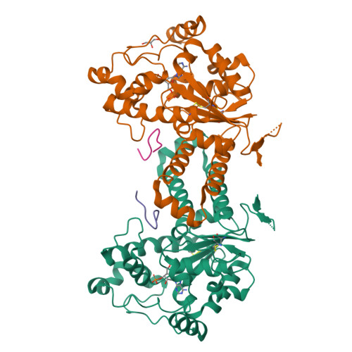
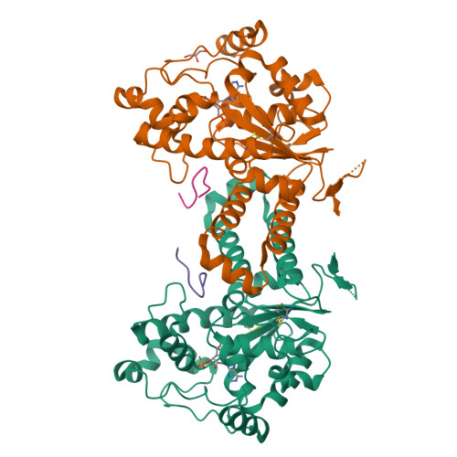
Crystal structure of human tyrosylprotein sulfotransferase-2 complexed with PAP and C4 peptide


Experimental Data Snapshot

  • Method: X-RAY DIFFRACTION
  • Resolution: 1.90 Å
  • R-Value Free: 0.222 
  • R-Value Work: 0.195 
  • R-Value Observed: 0.197 

wwPDB Validation   3D Report Full Report


Ligand Structure Quality Assessment 


This is version 1.2 of the entry. See complete history


Literature

Crystal structure of human tyrosylprotein sulfotransferase-2 reveals the mechanism of protein tyrosine sulfation reaction.

Teramoto, T.Fujikawa, Y.Kawaguchi, Y.Kurogi, K.Soejima, M.Adachi, R.Nakanishi, Y.Mishiro-Sato, E.Liu, M.C.Sakakibara, Y.Suiko, M.Kimura, M.Kakuta, Y.

(2013) Nat Commun 4: 1572-1572

  • DOI: https://doi.org/10.1038/ncomms2593
  • Primary Citation of Related Structures:  
    3AP1, 3AP3

  • PubMed Abstract: 

    Post-translational protein modification by tyrosine sulfation has an important role in extracellular protein-protein interactions. The protein tyrosine sulfation reaction is catalysed by the Golgi enzyme called the tyrosylprotein sulfotransferase. To date, no crystal structure is available for tyrosylprotein sulfotransferase. Detailed mechanism of protein tyrosine sulfation reaction has thus remained unclear. Here we present the first crystal structure of the human tyrosylprotein sulfotransferase isoform 2 complexed with a substrate peptide (C4P5Y3) derived from complement C4 and 3'-phosphoadenosine-5'-phosphate at 1.9 Å resolution. Structural and complementary mutational analyses revealed the molecular basis for catalysis being an SN2-like in-line displacement mechanism. Tyrosylprotein sulfotransferase isoform 2 appeared to recognize the C4 peptide in a deep cleft by using a short parallel β-sheet type interaction, and the bound C4P5Y3 forms an L-shaped structure. Surprisingly, the mode of substrate peptide recognition observed in the tyrosylprotein sulfotransferase isoform 2 structure resembles that observed for the receptor type tyrosine kinases.


  • Organizational Affiliation

    Laboratory of Structural Biology, Graduate School of Systems Life Sciences, Kyushu University, Hakozaki 6-10-1, Fukuoka 812-8581, Japan.


Macromolecules
Find similar proteins by:  (by identity cutoff)  |  3D Structure
Entity ID: 1
MoleculeChains Sequence LengthOrganismDetailsImage
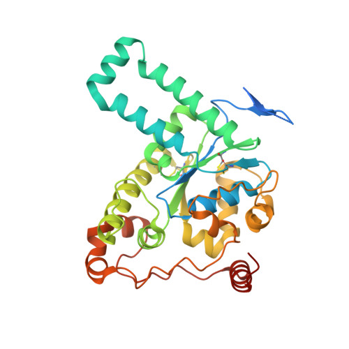
Protein-tyrosine sulfotransferase 2
A, B
337Homo sapiensMutation(s): 0 
Gene Names: TPST2
EC: 2.8.2.20
UniProt & NIH Common Fund Data Resources
Find proteins for O60704 (Homo sapiens)
Explore O60704 
Go to UniProtKB:  O60704
PHAROS:  O60704
GTEx:  ENSG00000128294 
Entity Groups  
Sequence Clusters30% Identity50% Identity70% Identity90% Identity95% Identity100% Identity
UniProt GroupO60704
Sequence Annotations
Expand
  • Reference Sequence

Find similar proteins by:  Sequence   |   3D Structure  

Entity ID: 2
MoleculeChains Sequence LengthOrganismDetailsImage
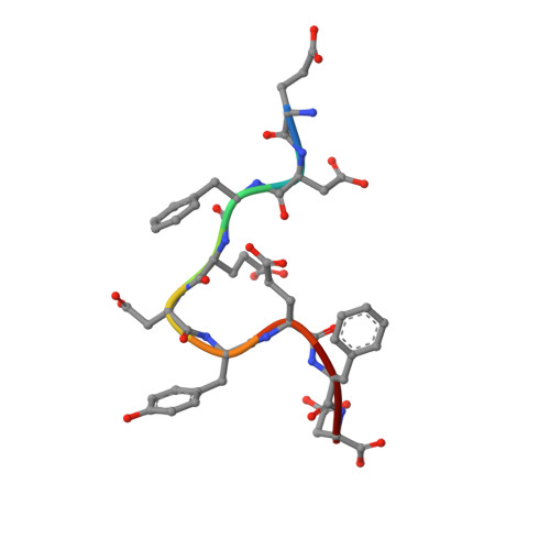
C4 peptideC [auth S],
D [auth T]
9N/AMutation(s): 0 
Sequence Annotations
Expand
  • Reference Sequence
Experimental Data & Validation

Experimental Data

  • Method: X-RAY DIFFRACTION
  • Resolution: 1.90 Å
  • R-Value Free: 0.222 
  • R-Value Work: 0.195 
  • R-Value Observed: 0.197 
  • Space Group: H 3 2
Unit Cell:
Length ( Å )Angle ( ˚ )
a = 138.263α = 90
b = 138.263β = 90
c = 227.994γ = 120
Software Package:
Software NamePurpose
ADSCdata collection
SHELXSphasing
REFMACrefinement
HKL-2000data reduction
HKL-2000data scaling

Structure Validation

View Full Validation Report


Ligand Structure Quality Assessment 


Entry History 

Revision History  (Full details and data files)

  • Version 1.0: 2011-10-26
    Type: Initial release
  • Version 1.1: 2013-03-27
    Changes: Database references
  • Version 1.2: 2024-10-16
    Changes: Data collection, Database references, Derived calculations, Structure summary