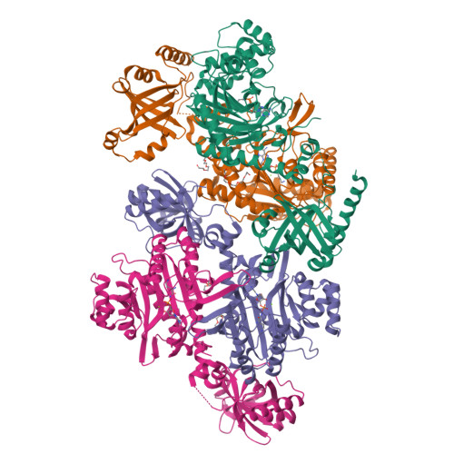
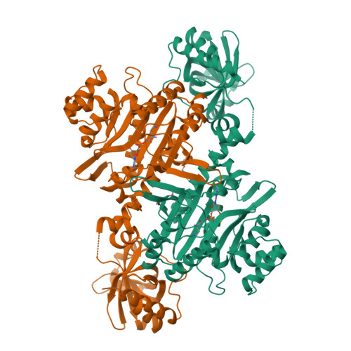
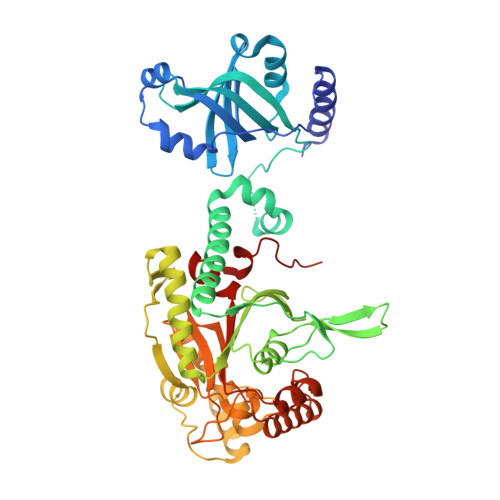
3E9I

Lysyl-tRNA synthetase from Bacillus stearothermophilus complexed with L-Lysine hydroxamate-AMP


Experimental Data Snapshot

  • Method: X-RAY DIFFRACTION
  • Resolution: 2.20 Å
  • R-Value Free: 0.246 
  • R-Value Work: 0.182 
  • R-Value Observed: 0.182 

wwPDB Validation   3D Report Full Report


Ligand Structure Quality Assessment 


This is version 1.2 of the entry. See complete history


Literature

Two crystal structures of lysyl-tRNA synthetase from Bacillus stearothermophilus in complex with lysyladenylate-like compounds: insights into the irreversible formation of the enzyme-bound adenylate of L-lysine hydroxamate

Sakurama, H.Takita, T.Mikami, B.Itoh, T.Yasukawa, K.Inouye, K.

(2009) J Biochem 145: 555-563

  • DOI: https://doi.org/10.1093/jb/mvp014
  • Primary Citation of Related Structures:  
    3E9H, 3E9I

  • PubMed Abstract: 

    Aminoacyl-tRNA synthetase forms an enzyme-bound intermediate, aminoacyladenylate in the amino-acid activation reaction. This reaction is monitored by measuring the ATP-PPi exchange reason in which [(32)P]PPi is incorporated into ATP. We previously reported that L-lysine hydroxamate completely inhibited the L-lysine-dependent ATP-PPi exchange reaction catalysed by lysyl-tRNA synthetase from Bacillus stearothermophilus (BsLysRS). Several experiments suggested that BsLysRS can adenylate L-lysine hydroxamate, but the enzyme-bound lysyladenylate-like compound does not undergo the nucleophilic attack of PPi. This contrasts with the two reports for seryl-tRNA synthetase (SerRS): (i) L-serine hydroxamate was utilized by yeast SerRS as a substrate in the ATP-PPi exchange; and (ii) a seryladenylate-like compound was formed from L-serine hydroxamate in the crystal structure of Thermus thermophilus SerRS. To gain clues about the mechanistic difference, we have determined the crystal structures of two complexes of BsLysRS with the adenylate of L-lysine hydroxamate and with 5'-O-[N-(L-Lysyl)sulphamoyl] adenosine. The comparisons of the two BsLysRS structures and the above SerRS structure revealed the specific side-chain shift of Glu411 of BsLysRS in the complex with the adenylate of L-lysine hydroxamate. In support of other structural comparisons, the result suggested that Glu411 plays a key role in the arrangement of PPi for the nucleophilic attack.


  • Organizational Affiliation

    Division of Food Science and Biotechnology, Graduate School of Agriculture, Kyoto University, Kyoto, Japan.


Macromolecules
Find similar proteins by:  (by identity cutoff)  |  3D Structure
Entity ID: 1
MoleculeChains Sequence LengthOrganismDetailsImage
Lysyl-tRNA synthetase
A, B, C, D
493Geobacillus stearothermophilusMutation(s): 0 
EC: 6.1.1.6
UniProt
Find proteins for Q9RHV9 (Geobacillus stearothermophilus)
Explore Q9RHV9 
Go to UniProtKB:  Q9RHV9
Entity Groups  
Sequence Clusters30% Identity50% Identity70% Identity90% Identity95% Identity100% Identity
UniProt GroupQ9RHV9
Sequence Annotations
Expand
  • Reference Sequence
Small Molecules
Ligands 3 Unique
IDChains Name / Formula / InChI Key2D Diagram3D Interactions
XAH
Query on XAH

Download Ideal Coordinates CCD File 
F [auth A],
I [auth B],
L [auth C],
P [auth D]
5'-O-{(R)-hydroxy[(L-lysylamino)oxy]phosphoryl}adenosine
C16 H27 N8 O8 P
UZJSLDPAROCUBS-LEJQEAHTSA-N
1PE
Query on 1PE

Download Ideal Coordinates CCD File 
G [auth A],
J [auth B],
M [auth C],
N [auth C]
PENTAETHYLENE GLYCOL
C10 H22 O6
JLFNLZLINWHATN-UHFFFAOYSA-N
MG
Query on MG

Download Ideal Coordinates CCD File 
E [auth A],
H [auth B],
K [auth C],
O [auth D]
MAGNESIUM ION
Mg
JLVVSXFLKOJNIY-UHFFFAOYSA-N
Binding Affinity Annotations 
IDSourceBinding Affinity
XAH PDBBind:  3E9I Ki: 600 (nM) from 1 assay(s)
Experimental Data & Validation

Experimental Data

  • Method: X-RAY DIFFRACTION
  • Resolution: 2.20 Å
  • R-Value Free: 0.246 
  • R-Value Work: 0.182 
  • R-Value Observed: 0.182 
  • Space Group: P 1 21 1
Unit Cell:
Length ( Å )Angle ( ˚ )
a = 79.143α = 90
b = 82.483β = 89.99
c = 149.779γ = 90
Software Package:
Software NamePurpose
CNSrefinement
HKL-2000data collection
HKL-2000data reduction
HKL-2000data scaling
CNSphasing

Structure Validation

View Full Validation Report


Ligand Structure Quality Assessment 


Entry History 

Deposition Data

Revision History  (Full details and data files)

  • Version 1.0: 2009-07-14
    Type: Initial release
  • Version 1.1: 2011-07-13
    Changes: Version format compliance
  • Version 1.2: 2024-03-20
    Changes: Data collection, Database references, Derived calculations