Structure of an odorant-binding protein from the mosquito Aedes aegypti suggests a binding pocket covered by a pH-sensitive "Lid".
Leite, N.R., Krogh, R., Xu, W., Ishida, Y., Iulek, J., Leal, W.S., Oliva, G.(2009) PLoS One 4: e8006-e8006
- PubMed: 19956631
- DOI: https://doi.org/10.1371/journal.pone.0008006
- Primary Citation of Related Structures:
3K1E - PubMed Abstract:
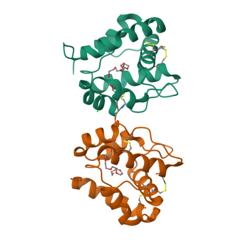
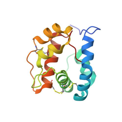
The yellow fever mosquito, Aedes aegypti, is the primary vector for the viruses that cause yellow fever, mostly in tropical regions of Africa and in parts of South America, and human dengue, which infects 100 million people yearly in the tropics and subtropics. A better understanding of the structural biology of olfactory proteins may pave the way for the development of environmentally-friendly mosquito attractants and repellents, which may ultimately contribute to reduction of mosquito biting and disease transmission. Previously, we isolated and cloned a major, female-enriched odorant-binding protein (OBP) from the yellow fever mosquito, AaegOBP1, which was later inadvertently renamed AaegOBP39. We prepared recombinant samples of AaegOBP1 by using an expression system that allows proper formation of disulfide bridges and generates functional OBPs, which are indistinguishable from native OBPs. We crystallized AaegOBP1 and determined its three-dimensional structure at 1.85 A resolution by molecular replacement based on the structure of the malaria mosquito OBP, AgamOBP1, the only mosquito OBP structure known to date. The structure of AaegOBP1 ( = AaegOBP39) shares the common fold of insect OBPs with six alpha-helices knitted by three disulfide bonds. A long molecule of polyethylene glycol (PEG) was built into the electron-density maps identified in a long tunnel formed by a crystallographic dimer of AaegOBP1. Circular dichroism analysis indicated that delipidated AaegOBP1 undergoes a pH-dependent conformational change, which may lead to release of odorant at low pH (as in the environment in the vicinity of odorant receptors). A C-terminal loop covers the binding cavity and this "lid" may be opened by disruption of an array of acid-labile hydrogen bonds thus explaining reduced or no binding affinity at low pH.
Organizational Affiliation:
Centro de Biotecnologia Molecular Estrutural, Instituto de Física de São Carlos, Universidade de São Paulo, São Carlos, São Paulo, Brazil.


