3WE0 | pdb_00003we0

L-Amino acid oxidase/monooxygenase from Pseudomonas sp. AIU 813


Experimental Data Snapshot

  • Method: X-RAY DIFFRACTION
  • Resolution: 1.90 Å
  • R-Value Free: 
    0.254 (Depositor), 0.250 (DCC) 
  • R-Value Work: 
    0.209 (Depositor), 0.210 (DCC) 
  • R-Value Observed: 
    0.212 (Depositor) 

wwPDB Validation   3D Report Full Report


Ligand Structure Quality Assessment 

Created with Raphaël 2.3.0Worse 01 BetterLigand structure goodness of fit to experimental dataBest fitted FADClick on this verticalbar to view details

This is version 1.2 of the entry. See complete history


Literature

Mutational and crystallographic analysis of l-amino acid oxidase/monooxygenase from Pseudomonas sp. AIU 813: Interconversion between oxidase and monooxygenase activities

Matsui, D.Im, D.H.Sugawara, A.Fukuta, Y.Fushinobu, S.Isobe, K.Asano, Y.

(2014) FEBS Open Bio 4: 220-228

  • DOI: https://doi.org/10.1016/j.fob.2014.02.002
  • Primary Citation of Related Structures:  
    3WE0

  • PubMed Abstract: 

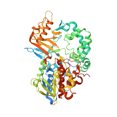
    In this study, it was shown for the first time that l-amino acid oxidase of Pseudomonas sp. AIU813, renamed as l-amino acid oxidase/monooxygenase (l-AAO/MOG), exhibits l-lysine 2-monooxygenase as well as oxidase activity. l-Lysine oxidase activity of l-AAO/MOG was increased in a p-chloromercuribenzoate (p-CMB) concentration-dependent manner to a final level that was five fold higher than that of the non-treated enzyme. In order to explain the effects of modification by the sulfhydryl reagent, saturation mutagenesis studies were carried out on five cysteine residues, and we succeeded in identifying l-AAO/MOG C254I mutant enzyme, which showed five-times higher specific activity of oxidase activity than that of wild type. The monooxygenase activity shown by the C254I variant was decreased significantly. Moreover, we also determined a high-resolution three-dimensional structure of l-AAO/MOG to provide a structural basis for its biochemical characteristics. The key residue for the activity conversion of l-AAO/MOG, Cys-254, is located near the aromatic cage (Trp-418, Phe-473, and Trp-516). Although the location of Cys-254 indicates that it is not directly involved in the substrate binding, the chemical modification by p-CMB or C254I mutation would have a significant impact on the substrate binding via the side chain of Trp-516. It is suggested that a slight difference of the binding position of a substrate can dictate the activity of this type of enzyme as oxidase or monooxygenase.


  • Organizational Affiliation

    Biotechnology Research Center and Department of Biotechnology, Toyama Prefectural University, 5180 Kurokawa, Imizu, Toyama 939-0398, Japan ; Asano Active Enzyme Molecule Project, ERATO, JST, 5180 Kurokawa, Imizu,Toyama 939-0398, Japan.


Macromolecules
Find similar proteins by:  (by identity cutoff)  |  3D Structure
Entity ID: 1
MoleculeChains Sequence LengthOrganismDetailsImage
L-amino acid oxidase/monooxygenase
A, B
580Pseudomonas sp.Mutation(s): 0 
Gene Names: laao
EC: 1.4.3.2 (PDB Primary Data), 1.13.12.3 (UniProt)
UniProt
Find proteins for B3IVI6 (Pseudomonas putida)
Explore B3IVI6 
Go to UniProtKB:  B3IVI6
Entity Groups  
Sequence Clusters30% Identity50% Identity70% Identity90% Identity95% Identity100% Identity
UniProt GroupB3IVI6
Sequence Annotations
Expand
  • Reference Sequence
Experimental Data & Validation

Experimental Data

  • Method: X-RAY DIFFRACTION
  • Resolution: 1.90 Å
  • R-Value Free:  0.254 (Depositor), 0.250 (DCC) 
  • R-Value Work:  0.209 (Depositor), 0.210 (DCC) 
  • R-Value Observed: 0.212 (Depositor) 
Space Group: P 21 21 2
Unit Cell:
Length ( Å )Angle ( ˚ )
a = 118.926α = 90
b = 141.353β = 90
c = 75.97γ = 90
Software Package:
Software NamePurpose
HKL-2000data collection
SOLVEphasing
REFMACrefinement
HKL-2000data reduction
HKL-2000data scaling

Structure Validation

View Full Validation Report


Ligand Structure Quality Assessment 

Created with Raphaël 2.3.0Worse 01 BetterLigand structure goodness of fit to experimental dataBest fitted FADClick on this verticalbar to view details

Entry History 

Deposition Data

Revision History  (Full details and data files)

  • Version 1.0: 2014-02-12
    Type: Initial release
  • Version 1.1: 2014-07-02
    Changes: Database references
  • Version 1.2: 2024-03-20
    Changes: Data collection, Database references, Derived calculations