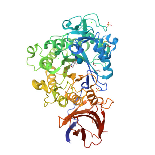
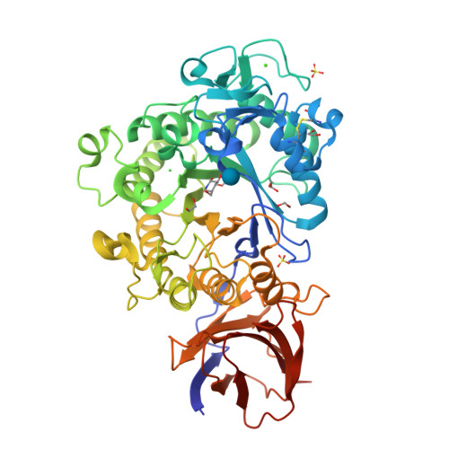
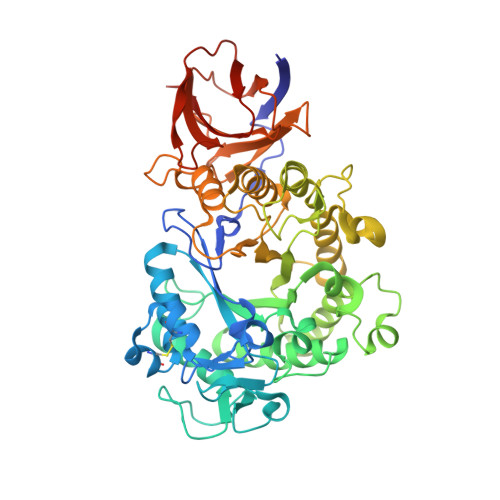
3ZQ9

Structure of a Paenibacillus Polymyxa Xyloglucanase from Glycoside Hydrolase Family 44


Experimental Data Snapshot

  • Method: X-RAY DIFFRACTION
  • Resolution: 1.86 Å
  • R-Value Free: 0.171 
  • R-Value Work: 0.137 
  • R-Value Observed: 0.138 

Starting Model: experimental
View more details

wwPDB Validation   3D Report Full Report


Ligand Structure Quality Assessment 


This is version 1.5 of the entry. See complete history


Literature

Structure and Activity of Paenibacillus Polymyxa Xyloglucanase from Glycoside Hydrolase Family 44.

Ariza, A.Eklof, J.M.Spadiut, O.Offen, W.A.Roberts, S.M.Besenmatter, W.Friis, E.P.Skjot, M.Wilson, K.S.Brumer, H.Davies, G.

(2011) J Biol Chem 286: 33890

  • DOI: https://doi.org/10.1074/jbc.M111.262345
  • Primary Citation of Related Structures:  
    2YIH, 2YJQ, 2YKK, 3ZQ9

  • PubMed Abstract: 

    The enzymatic degradation of plant polysaccharides is emerging as one of the key environmental goals of the early 21st century, impacting on many processes in the textile and detergent industries as well as biomass conversion to biofuels. One of the well known problems with the use of nonstarch (nonfood)-based substrates such as the plant cell wall is that the cellulose fibers are embedded in a network of diverse polysaccharides, including xyloglucan, that renders access difficult. There is therefore increasing interest in the "accessory enzymes," including xyloglucanases, that may aid biomass degradation through removal of "hemicellulose" polysaccharides. Here, we report the biochemical characterization of the endo-β-1,4-(xylo)glucan hydrolase from Paenibacillus polymyxa with polymeric, oligomeric, and defined chromogenic aryl-oligosaccharide substrates. The enzyme displays an unusual specificity on defined xyloglucan oligosaccharides, cleaving the XXXG-XXXG repeat into XXX and GXXXG. Kinetic analysis on defined oligosaccharides and on aryl-glycosides suggests that both the -4 and +1 subsites show discrimination against xylose-appended glucosides. The three-dimensional structures of PpXG44 have been solved both in apo-form and as a series of ligand complexes that map the -3 to -1 and +1 to +5 subsites of the extended ligand binding cleft. Complex structures are consistent with partial intolerance of xylosides in the -4' subsites. The atypical specificity of PpXG44 may thus find use in industrial processes involving xyloglucan degradation, such as biomass conversion, or in the emerging exciting applications of defined xyloglucans in food, pharmaceuticals, and cellulose fiber modification.


  • Organizational Affiliation

    Structural Biology Laboratory, Department of Chemistry, University of York, Heslington, York YO10 5DD, United Kingdom.


Macromolecules
Find similar proteins by:  (by identity cutoff)  |  3D Structure
Entity ID: 1
MoleculeChains Sequence LengthOrganismDetailsImage
XYLOGLUCANASE524Paenibacillus polymyxaMutation(s): 2 
EC: 3.2.1.4 (PDB Primary Data), 3.2.1.151 (PDB Primary Data)
UniProt
Find proteins for Q1A2D0 (Paenibacillus polymyxa)
Explore Q1A2D0 
Go to UniProtKB:  Q1A2D0
Entity Groups  
Sequence Clusters30% Identity50% Identity70% Identity90% Identity95% Identity100% Identity
UniProt GroupQ1A2D0
Sequence Annotations
Expand
  • Reference Sequence
Small Molecules
Ligands 6 Unique
IDChains Name / Formula / InChI Key2D Diagram3D Interactions
BGC
Query on BGC

Download Ideal Coordinates CCD File 
C [auth A]beta-D-glucopyranose
C6 H12 O6
WQZGKKKJIJFFOK-VFUOTHLCSA-N
NOY
Query on NOY

Download Ideal Coordinates CCD File 
B [auth A](2R,3S,4R,5R)-5-(HYDROXYMETHYL)PIPERIDINE-2,3,4-TRIOL
C6 H13 N O4
BHOYFRIRWXBNHP-ARQDHWQXSA-N
SO4
Query on SO4

Download Ideal Coordinates CCD File 
F [auth A],
G [auth A]
SULFATE ION
O4 S
QAOWNCQODCNURD-UHFFFAOYSA-L
EDO
Query on EDO

Download Ideal Coordinates CCD File 
H [auth A],
I [auth A],
J [auth A],
K [auth A]
1,2-ETHANEDIOL
C2 H6 O2
LYCAIKOWRPUZTN-UHFFFAOYSA-N
CA
Query on CA

Download Ideal Coordinates CCD File 
D [auth A]CALCIUM ION
Ca
BHPQYMZQTOCNFJ-UHFFFAOYSA-N
CL
Query on CL

Download Ideal Coordinates CCD File 
E [auth A]CHLORIDE ION
Cl
VEXZGXHMUGYJMC-UHFFFAOYSA-M
Experimental Data & Validation

Experimental Data

  • Method: X-RAY DIFFRACTION
  • Resolution: 1.86 Å
  • R-Value Free: 0.171 
  • R-Value Work: 0.137 
  • R-Value Observed: 0.138 
  • Space Group: P 31 2 1
Unit Cell:
Length ( Å )Angle ( ˚ )
a = 83.61α = 90
b = 83.61β = 90
c = 157.531γ = 120
Software Package:
Software NamePurpose
REFMACrefinement
MOSFLMdata reduction
SCALAdata scaling

Structure Validation

View Full Validation Report


Ligand Structure Quality Assessment 


Entry History 

Deposition Data

Revision History  (Full details and data files)

  • Version 1.0: 2011-06-22
    Type: Initial release
  • Version 1.1: 2011-07-27
    Changes: Other, Version format compliance
  • Version 1.2: 2012-04-25
    Changes: Other
  • Version 1.3: 2017-06-28
    Changes: Data collection
  • Version 1.4: 2020-07-29
    Type: Remediation
    Reason: Carbohydrate remediation
    Changes: Data collection, Derived calculations, Other, Structure summary
  • Version 1.5: 2023-12-20
    Changes: Data collection, Database references, Refinement description, Structure summary