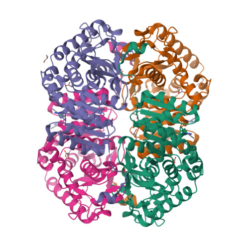
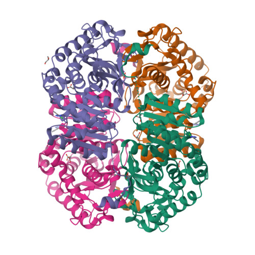
4AJ2

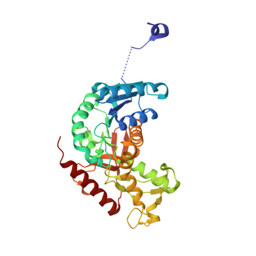
rat LDHA in complex with 5-(2-chlorophenyl)-1H-tetrazole


Experimental Data Snapshot

  • Method: X-RAY DIFFRACTION
  • Resolution: 1.75 Å
  • R-Value Free: 0.194 
  • R-Value Work: 0.158 
  • R-Value Observed: 0.160 

Starting Model: other
View more details

wwPDB Validation   3D Report Full Report


Ligand Structure Quality Assessment 


This is version 1.3 of the entry. See complete history


Literature

The Design and Synthesis of Novel Lactate Dehydrogenase a Inhibitors by Fragment-Based Lead Generation

Ward, R.Brassington, C.Breeze, A.L.Caputo, A.Critchlow, S.Davies, G.Goodwin, L.Hassall, G.Greenwood, R.Holdgate, G.Mrosek, M.Norman, R.A.Pearson, S.Tart, J.Tucker, J.A.Vogtherr, M.Whittaker, D.Wingfield, J.Winter, J.Hudson, K.

(2012) J Med Chem 55: 3285

  • DOI: https://doi.org/10.1021/jm201734r
  • Primary Citation of Related Structures:  
    4AJ1, 4AJ2, 4AJ4, 4AJE, 4AJH, 4AJI, 4AJJ, 4AJK, 4AJL, 4AJN, 4AJO, 4AJP, 4AL4

  • PubMed Abstract: 

    Lactate dehydrogenase A (LDHA) catalyzes the conversion of pyruvate to lactate, utilizing NADH as a cofactor. It has been identified as a potential therapeutic target in the area of cancer metabolism. In this manuscript we report our progress using fragment-based lead generation (FBLG), assisted by X-ray crystallography to develop small molecule LDHA inhibitors. Fragment hits were identified through NMR and SPR screening and optimized into lead compounds with nanomolar binding affinities via fragment linking. Also reported is their modification into cellular active compounds suitable for target validation work.


  • Organizational Affiliation

    Oncology and Discovery Sciences iMEDs, AstraZeneca, Mereside, Alderley Park, Macclesfield, Cheshire, SK10 4TG, UK. richard.a.ward@astrazeneca.com


Macromolecules
Find similar proteins by:  (by identity cutoff)  |  3D Structure
Entity ID: 1
MoleculeChains Sequence LengthOrganismDetailsImage
L-LACTATE DEHYDROGENASE A CHAIN
A, B, C, D
331Rattus norvegicusMutation(s): 0 
EC: 1.1.1.27
UniProt
Find proteins for P04642 (Rattus norvegicus)
Explore P04642 
Go to UniProtKB:  P04642
Entity Groups  
Sequence Clusters30% Identity50% Identity70% Identity90% Identity95% Identity100% Identity
UniProt GroupP04642
Sequence Annotations
Expand
  • Reference Sequence
Small Molecules
Ligands 3 Unique
IDChains Name / Formula / InChI Key2D Diagram3D Interactions
52C
Query on 52C

Download Ideal Coordinates CCD File 
F [auth A],
J [auth B],
N [auth C],
Q [auth D]
5-(2-CHLOROPHENYL)-1H-TETRAZOLE
C7 H5 Cl N4
PSUIIKIEUATWCZ-UHFFFAOYSA-N
MLI
Query on MLI

Download Ideal Coordinates CCD File 
G [auth A]
H [auth A]
K [auth B]
L [auth B]
O [auth C]
G [auth A],
H [auth A],
K [auth B],
L [auth B],
O [auth C],
P [auth C],
R [auth D],
S [auth D]
MALONATE ION
C3 H2 O4
OFOBLEOULBTSOW-UHFFFAOYSA-L
GOL
Query on GOL

Download Ideal Coordinates CCD File 
E [auth A],
I [auth B],
M [auth C]
GLYCEROL
C3 H8 O3
PEDCQBHIVMGVHV-UHFFFAOYSA-N
Binding Affinity Annotations 
IDSourceBinding Affinity
52C BindingDB:  4AJ2 Kd: 4.20e+6 (nM) from 1 assay(s)
IC50: 5.00e+5 (nM) from 1 assay(s)
PDBBind:  4AJ2 Kd: 4.20e+6 (nM) from 1 assay(s)
Experimental Data & Validation

Experimental Data

  • Method: X-RAY DIFFRACTION
  • Resolution: 1.75 Å
  • R-Value Free: 0.194 
  • R-Value Work: 0.158 
  • R-Value Observed: 0.160 
  • Space Group: P 1 21 1
Unit Cell:
Length ( Å )Angle ( ˚ )
a = 62α = 90
b = 81.73β = 96.02
c = 128.78γ = 90
Software Package:
Software NamePurpose
REFMACrefinement
d*TREKdata reduction
d*TREKdata scaling
AMoREphasing

Structure Validation

View Full Validation Report


Ligand Structure Quality Assessment 


Entry History 

Deposition Data

Revision History  (Full details and data files)

  • Version 1.0: 2012-03-21
    Type: Initial release
  • Version 1.1: 2012-04-25
    Changes: Other
  • Version 1.2: 2018-04-04
    Changes: Data collection
  • Version 1.3: 2024-05-01
    Changes: Data collection, Database references, Derived calculations, Other, Refinement description