4C69

ATP binding to murine voltage-dependent anion channel 1 (mVDAC1).


Experimental Data Snapshot

  • Method: X-RAY DIFFRACTION
  • Resolution: 2.28 Å
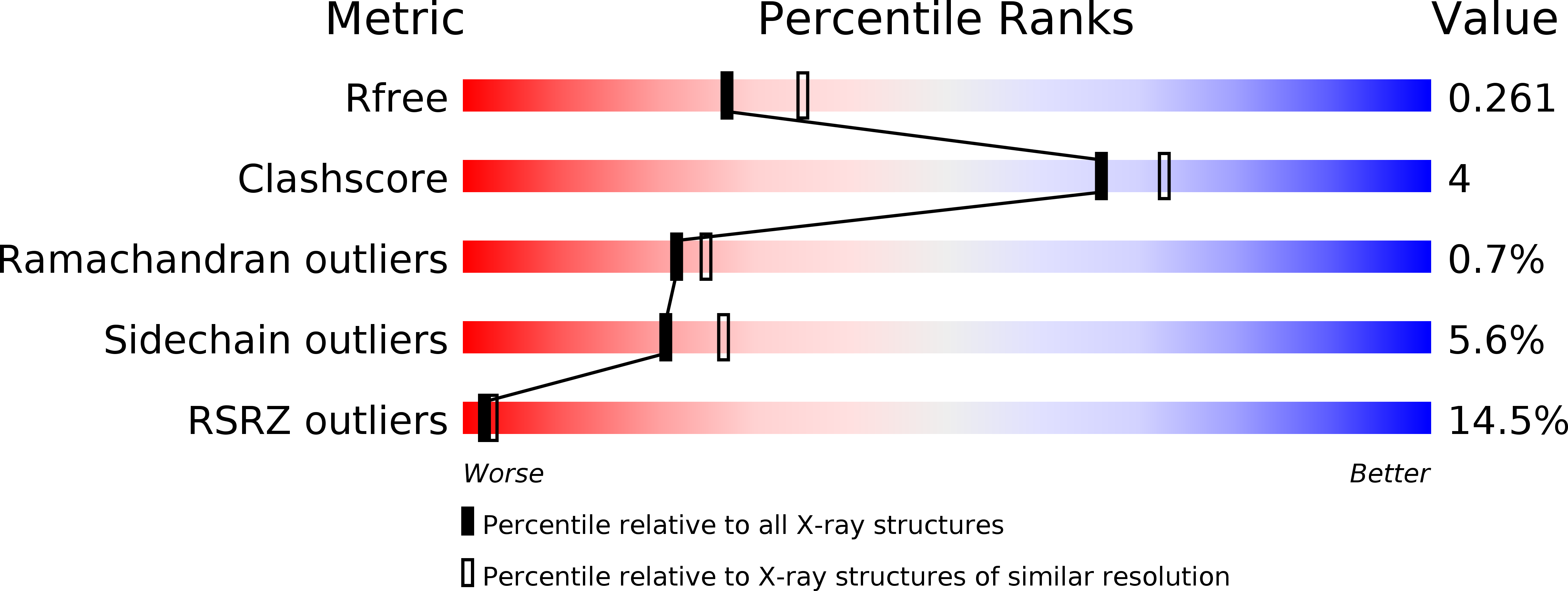
  • R-Value Free: 0.261 
  • R-Value Work: 0.204 
  • R-Value Observed: 0.207 

Starting Model: experimental
View more details

wwPDB Validation   3D Report Full Report


Ligand Structure Quality Assessment 


This is version 1.4 of the entry. See complete history


Literature

Structure-Guided Simulations Illuminate the Mechanism of ATP Transport Through Vdac1.

Choudhary, O.P.Paz, A.Adelman, J.L.Colletier, J.Abramson, J.Grabe, M.

(2014) Nat Struct Mol Biol 21: 626

  • DOI: https://doi.org/10.1038/nsmb.2841
  • Primary Citation of Related Structures:  
    4C69

  • PubMed Abstract: 

    The voltage-dependent anion channel (VDAC) mediates the flow of metabolites and ions across the outer mitochondrial membrane of all eukaryotic cells. The open channel passes millions of ATP molecules per second, whereas the closed state exhibits no detectable ATP flux. High-resolution structures of VDAC1 revealed a 19-stranded β-barrel with an α-helix partially occupying the central pore. To understand ATP permeation through VDAC, we solved the crystal structure of mouse VDAC1 (mVDAC1) in the presence of ATP, revealing a low-affinity binding site. Guided by these coordinates, we initiated hundreds of molecular dynamics simulations to construct a Markov state model of ATP permeation. These simulations indicate that ATP flows through VDAC through multiple pathways, in agreement with our structural data and experimentally determined physiological rates.


  • Organizational Affiliation

    1] Joint Carnegie Mellon University-University of Pittsburgh PhD Program in Computational Biology, Pittsburgh, Pennsylvania, USA. [2].


Macromolecules
Find similar proteins by:  (by identity cutoff)  |  3D Structure
Entity ID: 1
MoleculeChains Sequence LengthOrganismDetailsImage
VOLTAGE-DEPENDENT ANION-SELECTIVE CHANNEL PROTEIN 1A [auth X]295Mus musculusMutation(s): 0 
Membrane Entity: Yes 
UniProt & NIH Common Fund Data Resources
Find proteins for Q60932 (Mus musculus)
Explore Q60932 
Go to UniProtKB:  Q60932
IMPC:  MGI:106919
Entity Groups  
Sequence Clusters30% Identity50% Identity70% Identity90% Identity95% Identity100% Identity
UniProt GroupQ60932
Sequence Annotations
Expand
  • Reference Sequence
Small Molecules
Ligands 3 Unique
IDChains Name / Formula / InChI Key2D Diagram3D Interactions
MC3
Query on MC3

Download Ideal Coordinates CCD File 
C [auth X],
F [auth X]
1,2-DIMYRISTOYL-RAC-GLYCERO-3-PHOSPHOCHOLINE
C36 H72 N O8 P
CITHEXJVPOWHKC-UUWRZZSWSA-N
ATP
Query on ATP

Download Ideal Coordinates CCD File 
E [auth X]ADENOSINE-5'-TRIPHOSPHATE
C10 H16 N5 O13 P3
ZKHQWZAMYRWXGA-KQYNXXCUSA-N
LDA
Query on LDA

Download Ideal Coordinates CCD File 
B [auth X]
D [auth X]
G [auth X]
H [auth X]
I [auth X]
B [auth X],
D [auth X],
G [auth X],
H [auth X],
I [auth X],
J [auth X]
LAURYL DIMETHYLAMINE-N-OXIDE
C14 H31 N O
SYELZBGXAIXKHU-UHFFFAOYSA-N
Experimental Data & Validation

Experimental Data

  • Method: X-RAY DIFFRACTION
  • Resolution: 2.28 Å
  • R-Value Free: 0.261 
  • R-Value Work: 0.204 
  • R-Value Observed: 0.207 
  • Space Group: C 1 2 1
Unit Cell:
Length ( Å )Angle ( ˚ )
a = 100.152α = 90
b = 58.369β = 99.25
c = 66.545γ = 90
Software Package:
Software NamePurpose
PHENIXrefinement
XDSdata reduction
XSCALEdata scaling
PHENIXphasing

Structure Validation

View Full Validation Report


Ligand Structure Quality Assessment 


Entry History 

Deposition Data

Revision History  (Full details and data files)

  • Version 1.0: 2014-06-04
    Type: Initial release
  • Version 1.1: 2014-06-11
    Changes: Database references
  • Version 1.2: 2014-06-18
    Changes: Database references
  • Version 1.3: 2014-07-16
    Changes: Database references
  • Version 1.4: 2023-12-20
    Changes: Data collection, Database references, Derived calculations, Other, Refinement description