4IWQ

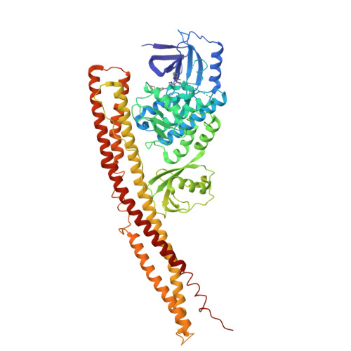
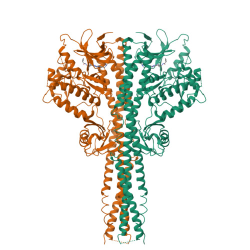
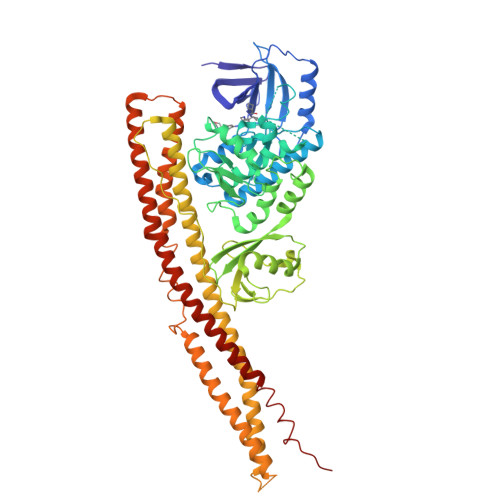
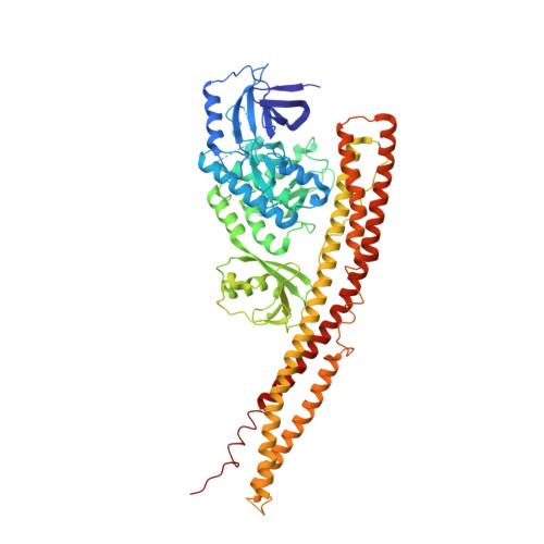
Crystal structure and mechanism of activation of TBK1


Experimental Data Snapshot

  • Method: X-RAY DIFFRACTION
  • Resolution: 3.00 Å
  • R-Value Free: 0.310 
  • R-Value Work: 0.262 
  • R-Value Observed: 0.264 

wwPDB Validation   3D Report Full Report


Ligand Structure Quality Assessment 


This is version 1.2 of the entry. See complete history


Literature

Crystal structure and mechanism of activation of TANK-binding kinase 1.

Larabi, A.Devos, J.M.Ng, S.L.Nanao, M.H.Round, A.Maniatis, T.Panne, D.

(2013) Cell Rep 3: 734-746

  • DOI: https://doi.org/10.1016/j.celrep.2013.01.034
  • Primary Citation of Related Structures:  
    4IW0, 4IWO, 4IWP, 4IWQ

  • PubMed Abstract: 

    Tank-binding kinase I (TBK1) plays a key role in the innate immune system by integrating signals from pattern-recognition receptors. Here, we report the X-ray crystal structures of inhibitor-bound inactive and active TBK1 determined to 2.6 Å and 4.0 Å resolution, respectively. The structures reveal a compact dimer made up of trimodular subunits containing an N-terminal kinase domain (KD), a ubiquitin-like domain (ULD), and an α-helical scaffold dimerization domain (SDD). Activation rearranges the KD into an active conformation while maintaining the overall dimer conformation. Low-resolution SAXS studies reveal that the missing C-terminal domain (CTD) extends away from the main body of the kinase dimer. Mutants that interfere with TBK1 dimerization show significantly reduced trans-autophosphorylation but retain the ability to bind adaptor proteins through the CTD. Our results provide detailed insights into the architecture of TBK1 and the molecular mechanism of activation.


  • Organizational Affiliation

    EMBL Grenoble, 6 Rue Jules Horowitz, Grenoble 38042, France.


Macromolecules
Find similar proteins by:  (by identity cutoff)  |  3D Structure
Entity ID: 1
MoleculeChains Sequence LengthOrganismDetailsImage
Serine/threonine-protein kinase TBK1659Homo sapiensMutation(s): 1 
Gene Names: NAKTBK1
EC: 2.7.11.1
UniProt & NIH Common Fund Data Resources
Find proteins for Q9UHD2 (Homo sapiens)
Explore Q9UHD2 
Go to UniProtKB:  Q9UHD2
PHAROS:  Q9UHD2
GTEx:  ENSG00000183735 
Entity Groups  
Sequence Clusters30% Identity50% Identity70% Identity90% Identity95% Identity100% Identity
UniProt GroupQ9UHD2
Sequence Annotations
Expand
  • Reference Sequence
Small Molecules
Ligands 1 Unique
IDChains Name / Formula / InChI Key2D Diagram3D Interactions
1FV
Query on 1FV

Download Ideal Coordinates CCD File 
B [auth A]N-{3-[(5-cyclopropyl-2-{[3-(morpholin-4-ylmethyl)phenyl]amino}pyrimidin-4-yl)amino]propyl}cyclobutanecarboxamide
C26 H36 N6 O2
UKBGBACORPRCGG-UHFFFAOYSA-N
Experimental Data & Validation

Experimental Data

  • Method: X-RAY DIFFRACTION
  • Resolution: 3.00 Å
  • R-Value Free: 0.310 
  • R-Value Work: 0.262 
  • R-Value Observed: 0.264 
  • Space Group: P 32 2 1
Unit Cell:
Length ( Å )Angle ( ˚ )
a = 135.76α = 90
b = 135.76β = 90
c = 86.5γ = 120
Software Package:
Software NamePurpose
PHENIXrefinement
PDB_EXTRACTdata extraction
ADSCdata collection
XDSdata reduction
XDSdata scaling
PHASERphasing

Structure Validation

View Full Validation Report


Ligand Structure Quality Assessment 


Entry History 

Deposition Data

Revision History  (Full details and data files)

  • Version 1.0: 2013-03-13
    Type: Initial release
  • Version 1.1: 2013-05-22
    Changes: Database references
  • Version 1.2: 2024-02-28
    Changes: Data collection, Database references, Derived calculations