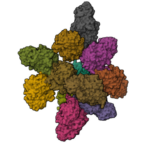
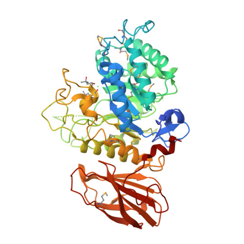
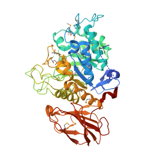
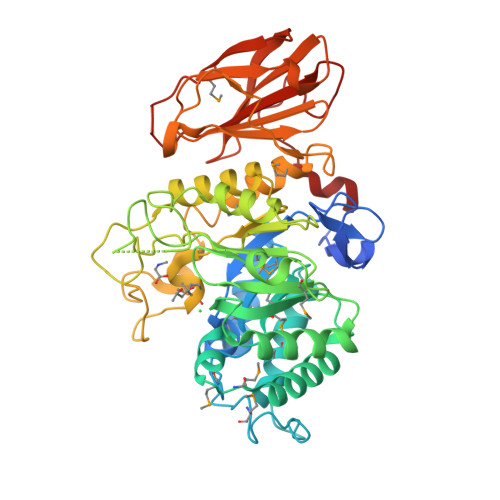
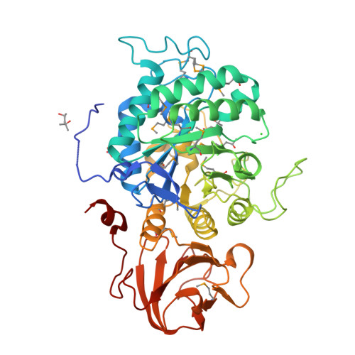
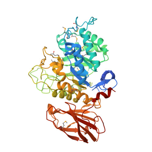
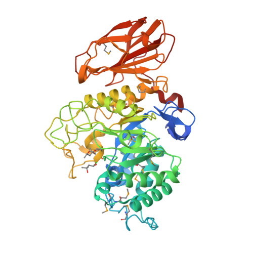
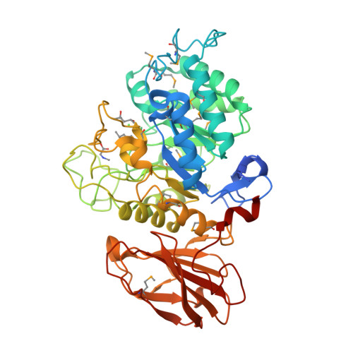
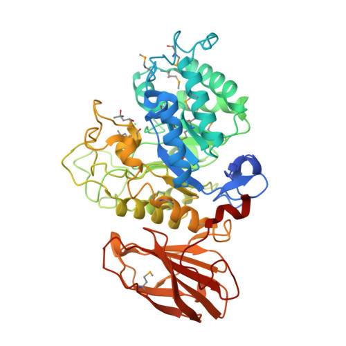
Crystal structure of a glycoside hydrolase family 5 (BVU_2644) from Bacteroides vulgatus ATCC 8482 at 1.90 A resolution
Joint Center for Structural Genomics (JCSG)To be published.
Experimental Data Snapshot
wwPDB Validation 3D Report Full Report

Entity ID: 1 | |||||
|---|---|---|---|---|---|
| Molecule | Chains | Sequence Length | Organism | Details | Image |
| glycoside hydrolase family 5 | 484 | Phocaeicola vulgatus ATCC 8482 | Mutation(s): 0 Gene Names: BVU_2644 | 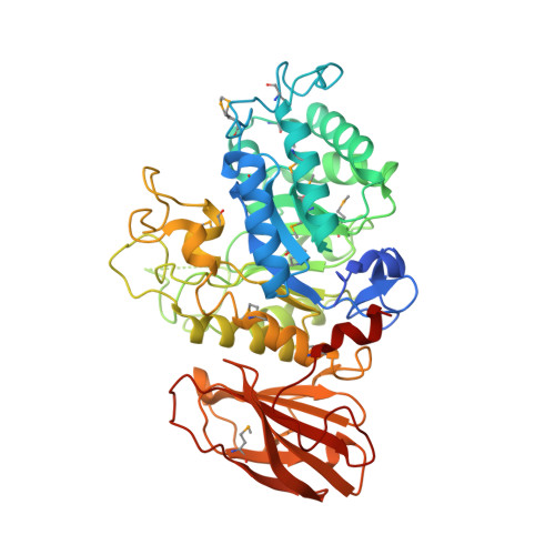 | |
UniProt | |||||
Find proteins for A6L3N2 (Phocaeicola vulgatus (strain ATCC 8482 / DSM 1447 / JCM 5826 / CCUG 4940 / NBRC 14291 / NCTC 11154)) Explore A6L3N2 Go to UniProtKB: A6L3N2 | |||||
Entity Groups | |||||
| Sequence Clusters | 30% Identity50% Identity70% Identity90% Identity95% Identity100% Identity | ||||
| UniProt Group | A6L3N2 | ||||
Sequence AnnotationsExpand | |||||
| |||||
| Ligands 4 Unique | |||||
|---|---|---|---|---|---|
| ID | Chains | Name / Formula / InChI Key | 2D Diagram | 3D Interactions | |
| MRD Query on MRD | AA [auth E] AB [auth L] BA [auth E] EA [auth F] FA [auth F] | (4R)-2-METHYLPENTANE-2,4-DIOL C6 H14 O2 SVTBMSDMJJWYQN-RXMQYKEDSA-N |  | ||
| SAR Query on SAR | KA [auth G], U [auth C], XA [auth K] | SARCOSINE C3 H7 N O2 FSYKKLYZXJSNPZ-UHFFFAOYSA-N |  | ||
| NO3 Query on NO3 | CA [auth E], GA [auth F], R [auth B] | NITRATE ION N O3 NHNBFGGVMKEFGY-UHFFFAOYSA-N |  | ||
| CL Query on CL | BB [auth L] DA [auth E] HA [auth F] LA [auth G] O [auth A] | CHLORIDE ION Cl VEXZGXHMUGYJMC-UHFFFAOYSA-M |  | ||
| Modified Residues 1 Unique | |||||
|---|---|---|---|---|---|
| ID | Chains | Type | Formula | 2D Diagram | Parent |
| MSE Query on MSE | A, B, C, D, E | L-PEPTIDE LINKING | C5 H11 N O2 Se |  | MET |
| Length ( Å ) | Angle ( ˚ ) |
|---|---|
| a = 115.95 | α = 91.6 |
| b = 117.589 | β = 92.82 |
| c = 125.249 | γ = 98.92 |
| Software Name | Purpose |
|---|---|
| MolProbity | model building |
| PDB_EXTRACT | data extraction |
| SHELX | phasing |
| SHARP | phasing |
| XSCALE | data scaling |
| BUSTER-TNT | refinement |
| PHASER | phasing |
| XDS | data reduction |
| SHELXD | phasing |
| autoSHARP | phasing |
| BUSTER | refinement |