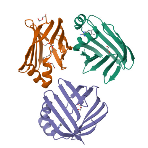
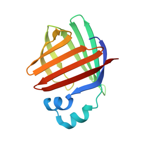
5L8N

crystal structure of human FABP6 protein with fragment 1


Experimental Data Snapshot

  • Method: X-RAY DIFFRACTION
  • Resolution: 2.12 Å
  • R-Value Free: 0.305 
  • R-Value Work: 0.247 
  • R-Value Observed: 0.249 

Starting Model: experimental
View more details

wwPDB Validation   3D Report Full Report


Ligand Structure Quality Assessment 


This is version 1.3 of the entry. See complete history


Literature

Identification and Investigation of Novel Binding Fragments in the Fatty Acid Binding Protein 6 (FABP6).

Hendrick, A.G.Muller, I.Willems, H.Leonard, P.M.Irving, S.Davenport, R.Ito, T.Reeves, J.Wright, S.Allen, V.Wilkinson, S.Heffron, H.Bazin, R.Turney, J.Mitchell, P.J.

(2016) J Med Chem 59: 8094-8102

  • DOI: https://doi.org/10.1021/acs.jmedchem.6b00869
  • Primary Citation of Related Structures:  
    5L8I, 5L8N, 5L8O

  • PubMed Abstract: 

    Fatty acid binding protein 6 (FABP6) is a potential drug discovery target, which, if inhibited, may have a therapeutic benefit for the treatment of diabetes. Currently, there are no published inhibitors of FABP6, and with the target believed to be amenable to fragment-based drug discovery, a structurally enabled program was initiated. This program successfully identified fragment hits using the surface plasmon resonance (SPR) platform. Several hits were validated with SAR and were found to be displaced by the natural ligand taurocholate. We report the first crystal structure of human FABP6 in the unbound form, in complex with cholate, and with one of the key fragments.


  • Organizational Affiliation

    Takeda Cambridge , 418 Cambridge Science Park, Cambridge CB4 0PZ, United Kingdom.


Macromolecules
Find similar proteins by:  (by identity cutoff)  |  3D Structure
Entity ID: 1
MoleculeChains Sequence LengthOrganismDetailsImage
Gastrotropin
A, B, C
128Homo sapiensMutation(s): 0 
Gene Names: FABP6ILBPILLBP
UniProt & NIH Common Fund Data Resources
Find proteins for P51161 (Homo sapiens)
Explore P51161 
Go to UniProtKB:  P51161
PHAROS:  P51161
GTEx:  ENSG00000170231 
Entity Groups  
Sequence Clusters30% Identity50% Identity70% Identity90% Identity95% Identity100% Identity
UniProt GroupP51161
Sequence Annotations
Expand
  • Reference Sequence
Small Molecules
Ligands 3 Unique
IDChains Name / Formula / InChI Key2D Diagram3D Interactions
P33
Query on P33

Download Ideal Coordinates CCD File 
H [auth B]3,6,9,12,15,18-HEXAOXAICOSANE-1,20-DIOL
C14 H30 O8
XPJRQAIZZQMSCM-UHFFFAOYSA-N
6RQ
Query on 6RQ

Download Ideal Coordinates CCD File 
D [auth A],
I [auth C]
5,6-dimethyl-1~{H}-benzimidazol-2-amine
C9 H11 N3
YPFQISHSXCFZMU-UHFFFAOYSA-N
PEG
Query on PEG

Download Ideal Coordinates CCD File 
E [auth A],
F [auth B],
G [auth B],
J [auth C]
DI(HYDROXYETHYL)ETHER
C4 H10 O3
MTHSVFCYNBDYFN-UHFFFAOYSA-N
Experimental Data & Validation

Experimental Data

  • Method: X-RAY DIFFRACTION
  • Resolution: 2.12 Å
  • R-Value Free: 0.305 
  • R-Value Work: 0.247 
  • R-Value Observed: 0.249 
  • Space Group: P 1 21 1
Unit Cell:
Length ( Å )Angle ( ˚ )
a = 47.25α = 90
b = 64.43β = 94.27
c = 59.02γ = 90
Software Package:
Software NamePurpose
REFMACrefinement
XDSdata reduction
Aimlessdata scaling

Structure Validation

View Full Validation Report


Ligand Structure Quality Assessment 


Entry History 

Deposition Data

Revision History  (Full details and data files)

  • Version 1.0: 2016-08-24
    Type: Initial release
  • Version 1.1: 2016-08-31
    Changes: Database references
  • Version 1.2: 2016-09-21
    Changes: Database references
  • Version 1.3: 2024-01-10
    Changes: Data collection, Database references, Refinement description, Structure summary