Mapping Hydrophobic Tunnels and Cavities in Neuroglobin with Noble Gas under Pressure.
Colloc'h, N., Carpentier, P., Montemiglio, L.C., Vallone, B., Prange, T.(2017) Biophys J 113: 2199-2206
- PubMed: 29108649
- DOI: https://doi.org/10.1016/j.bpj.2017.10.014
- Primary Citation of Related Structures:
5NVI, 5NW6, 5O17, 5O18, 5O1K, 5O27, 6EYE - PubMed Abstract:
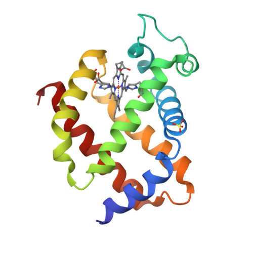
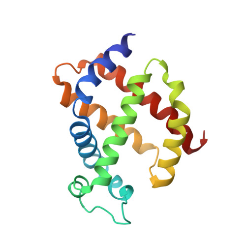
Internal cavities are crucial for conformational flexibility of proteins and can be mapped through noble gas diffusion and docking. Here we investigate the hydrophobic cavities and tunnel network in neuroglobin (Ngb), a hexacoordinated heme protein likely to be involved in neuroprotection, using crystallography under noble gas pressure, mostly at room temperature. In murine Ngb, a large internal cavity is involved in the heme sliding mechanism to achieve binding of gaseous ligands through coordination to the heme iron. In this study, we report that noble gases are hosted by two major sites within the internal cavity. We propose that these cavities could store oxygen and allow its relay in the heme proximity, which could correspond to NO location in the nitrite-reductase function of Ngb. Thanks to a recently designed pressurization cell using krypton at high pressure, a new gas binding site has been characterized that reveals an alternate pathway for gaseous ligands. A new gas binding site on the proximal side of the heme has also been characterized, using xenon pressure on a Ngb mutant (V140W) that binds CO with a similar rate and affinity to the wild-type, despite a reshaping of the internal cavity. Moreover, this study, to our knowledge, provides new insights into the determinants of the heme sliding mechanism, suggesting that the shift at the beginning of helix G precedes and drives this process.
Organizational Affiliation:
ISTCT CNRS UNICAEN CEA Normandie University, CERVOxy Team, Centre Cyceron, Caen, France. Electronic address: colloch@cyceron.fr.


