6J15 | pdb_00006j15

Complex structure of GY-5 Fab and PD-1


Experimental Data Snapshot

  • Method: X-RAY DIFFRACTION
  • Resolution: 2.60 Å
  • R-Value Free: 
    0.277 (Depositor), 0.280 (DCC) 
  • R-Value Work: 
    0.212 (Depositor), 0.210 (DCC) 
  • R-Value Observed: 
    0.215 (Depositor) 

Starting Models: experimental
View more details

wwPDB Validation   3D Report Full Report


Ligand Structure Quality Assessment 

Created with Raphaël 2.3.0Worse 01 BetterLigand structure goodness of fit to experimental dataBest fitted NAGClick on this verticalbar to view details

This is version 2.2 of the entry. See complete history


Literature

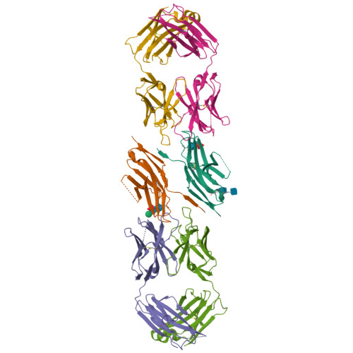
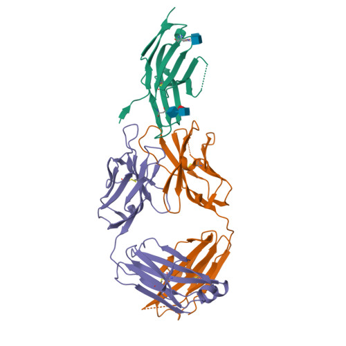
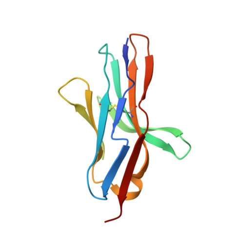
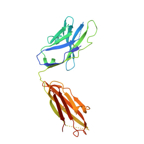
The FG Loop of PD-1 Serves as a "Hotspot" for Therapeutic Monoclonal Antibodies in Tumor Immune Checkpoint Therapy.

Chen, D.Tan, S.Zhang, H.Wang, H.He, W.Shi, R.Tong, Z.Zhu, J.Cheng, H.Gao, S.Chai, Y.Qi, J.Xiao, M.Yan, J.Gao, G.F.

(2019) iScience 14: 113-124

  • DOI: https://doi.org/10.1016/j.isci.2019.03.017
  • Primary Citation of Related Structures:  
    6J14, 6J15

  • PubMed Abstract: 

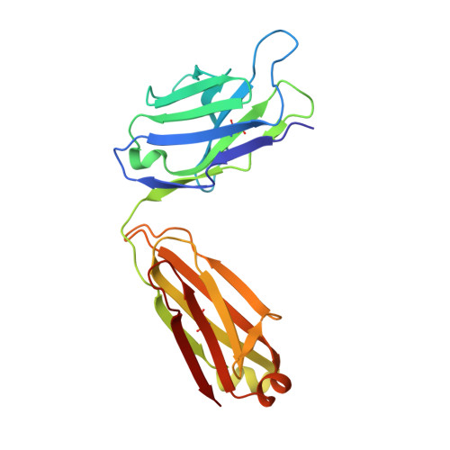
    Programmed cell death 1 (PD-1)/PD-1 ligand-1 (PD-L1)-blocking monoclonal antibodies (mAbs) have taken center stage for tumor immune checkpoint therapy. Identification of the "hotspots" on PD-1 for mAbs will help to develop next-generation oral deliverable agents with long-lasting efficacy. Here, we identified two PD-1-targeting mAbs, GY-5 and GY-14, with PD-1/PD-L1-blocking efficacy. Complex structural information revealed that both mAbs mainly bind to the FG loop of PD-1, which also contributes multiple interactions with PD-L1. The FG loop adopts substantially varied conformations upon binding to different mAbs, providing a novel targetable region for the development of PD-1-specific biologics and small chemical molecules. Glycosylation modifications of PD-1 could be observed in three of the four potential N-linked glycosylation sites. However, the binding of GY-5 and GY-14 to PD-1 was not affected by glycosylation. These findings broaden our understanding of the mechanism of anti-PD-1 mAbs and provide insight into the development of agents targeting PD-1.


  • Organizational Affiliation

    CAS Key Laboratory of Pathogenic Microbiology and Immunology, Institute of Microbiology, Chinese Academy of Sciences, Beijing 100101, China.


Macromolecules
Find similar proteins by:  (by identity cutoff)  |  3D Structure
Entity ID: 1
MoleculeChains Sequence LengthOrganismDetailsImage
Programmed cell death protein 1A [auth C],
B [auth D]
120Homo sapiensMutation(s): 0 
Gene Names: PDCD1PD1
UniProt & NIH Common Fund Data Resources
Find proteins for Q15116 (Homo sapiens)
Explore Q15116 
Go to UniProtKB:  Q15116
PHAROS:  Q15116
GTEx:  ENSG00000188389 
Entity Groups  
Sequence Clusters30% Identity50% Identity70% Identity90% Identity95% Identity100% Identity
UniProt GroupQ15116
Glycosylation
Glycosylation Sites: 3Go to GlyGen: Q15116-1
Sequence Annotations
Expand
  • Reference Sequence
Find similar proteins by:  (by identity cutoff)  |  3D Structure
Entity ID: 2
MoleculeChains Sequence LengthOrganismDetailsImage
GY-5 heavy chain FabC [auth H],
E [auth A]
219Mus musculusMutation(s): 0 
Entity Groups  
Sequence Clusters30% Identity50% Identity70% Identity90% Identity95% Identity100% Identity
Sequence Annotations
Expand
  • Reference Sequence
Find similar proteins by:  (by identity cutoff)  |  3D Structure
Entity ID: 3
MoleculeChains Sequence LengthOrganismDetailsImage
GY-5 light chain FabD [auth L],
F [auth B]
216Mus musculusMutation(s): 0 
Entity Groups  
Sequence Clusters30% Identity50% Identity70% Identity90% Identity95% Identity100% Identity
Sequence Annotations
Expand
  • Reference Sequence
Oligosaccharides

Help

Entity ID: 4
MoleculeChains Length2D Diagram Glycosylation3D Interactions
2-acetamido-2-deoxy-beta-D-glucopyranose-(1-4)-[alpha-L-fucopyranose-(1-6)]2-acetamido-2-deoxy-beta-D-glucopyranoseG [auth E]3N-Glycosylation
Glycosylation Resources
GlyTouCan:  G21290RB
GlyCosmos:  G21290RB
GlyGen:  G21290RB
Entity ID: 5
MoleculeChains Length2D Diagram Glycosylation3D Interactions
alpha-D-mannopyranose-(1-4)-2-acetamido-2-deoxy-beta-D-glucopyranose-(1-4)-[alpha-L-fucopyranose-(1-6)]2-acetamido-2-deoxy-beta-D-glucopyranoseH [auth F]4N-Glycosylation
Glycosylation Resources
GlyTouCan:  G47477HI
GlyCosmos:  G47477HI
GlyGen:  G47477HI
Experimental Data & Validation

Experimental Data

  • Method: X-RAY DIFFRACTION
  • Resolution: 2.60 Å
  • R-Value Free:  0.277 (Depositor), 0.280 (DCC) 
  • R-Value Work:  0.212 (Depositor), 0.210 (DCC) 
  • R-Value Observed: 0.215 (Depositor) 
Space Group: P 1 21 1
Unit Cell:
Length ( Å )Angle ( ˚ )
a = 61.595α = 90
b = 64.347β = 98.04
c = 151.481γ = 90
Software Package:
Software NamePurpose
PHENIXrefinement
HKL-3000data reduction
HKL-3000data scaling
PHASERphasing

Structure Validation

View Full Validation Report


Ligand Structure Quality Assessment 

Created with Raphaël 2.3.0Worse 01 BetterLigand structure goodness of fit to experimental dataBest fitted NAGClick on this verticalbar to view details

Entry History 

Deposition Data

Revision History  (Full details and data files)

  • Version 1.0: 2019-11-06
    Type: Initial release
  • Version 2.0: 2020-07-29
    Type: Remediation
    Reason: Carbohydrate remediation
    Changes: Atomic model, Data collection, Derived calculations, Structure summary
  • Version 2.1: 2023-11-22
    Changes: Data collection, Database references, Derived calculations, Refinement description, Structure summary
  • Version 2.2: 2024-11-13
    Changes: Structure summary