6JBT | pdb_00006jbt

Complex structure of toripalimab-Fab and PD-1


Experimental Data Snapshot

  • Method: X-RAY DIFFRACTION
  • Resolution: 2.47 Å
  • R-Value Free: 
    0.252 (Depositor), 0.250 (DCC) 
  • R-Value Work: 
    0.201 (Depositor), 0.200 (DCC) 
  • R-Value Observed: 
    0.203 (Depositor) 

Starting Model: experimental
View more details

wwPDB Validation   3D Report Full Report


Ligand Structure Quality Assessment 

Created with Raphaël 2.3.0Worse 01 BetterLigand structure goodness of fit to experimental dataBest fitted NAGClick on this verticalbar to view details

This is version 2.2 of the entry. See complete history


Literature

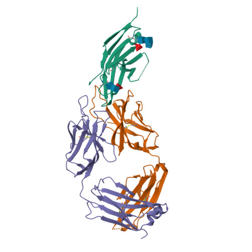
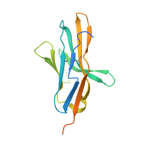
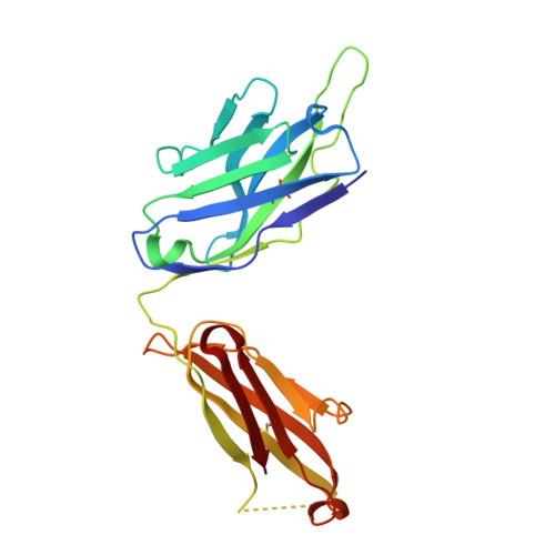
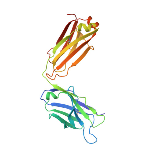
Glycosylation-independent binding of monoclonal antibody toripalimab to FG loop of PD-1 for tumor immune checkpoint therapy.

Liu, H.Guo, L.Zhang, J.Zhou, Y.Zhou, J.Yao, J.Wu, H.Yao, S.Chen, B.Chai, Y.Qi, J.Gao, G.F.Tan, S.Feng, H.Yan, J.

(2019) MAbs 11: 681-690

  • DOI: https://doi.org/10.1080/19420862.2019.1596513
  • Primary Citation of Related Structures:  
    6JBT

  • PubMed Abstract: 

    Monoclonal antibody (mAb)-based blockade of programmed cell death 1 (PD-1) or its ligand to enable antitumor T-cell immunity has been successful in treating multiple tumors. However, the structural basis of the binding mechanisms of the mAbs and PD-1 and the effects of glycosylation of PD-1 on mAb interaction are not well understood. Here, we report the complex structure of PD-1 with toripalimab, a mAb that is approved by China National Medical Products Administration as a second-line treatment for melanoma and is under multiple Phase 1-Phase 3 clinical trials in both China and the US. Our analysis reveals that toripalimab mainly binds to the FG loop of PD-1 with an unconventionally long complementarity-determining region 3 loop of the heavy chain, which is distinct from the known binding epitopes of anti-PD-1 mAbs with structural evidences. The glycan modifications of PD-1 could be observed in three potential N-linked glycosylation sites, while no substantial influences were detected to the binding of toripalimab. These findings benefit our understanding of the binding mechanisms of toripalimab to PD-1 and shed light for future development of biologics targeting PD-1. Atomic coordinates have been deposited in the Protein Data Bank under accession code 6JBT.


  • Organizational Affiliation

    a Department of Antibody Discovery and Engineering , Shanghai Junshi Biosciences Co., Ltd , Shanghai , China.


Macromolecules
Find similar proteins by:  (by identity cutoff)  |  3D Structure
Entity ID: 1
MoleculeChains Sequence LengthOrganismDetailsImage
Programmed cell death protein 1A [auth F]157Homo sapiensMutation(s): 0 
Gene Names: PDCD1PD1
UniProt & NIH Common Fund Data Resources
Find proteins for Q15116 (Homo sapiens)
Explore Q15116 
Go to UniProtKB:  Q15116
PHAROS:  Q15116
GTEx:  ENSG00000188389 
Entity Groups  
Sequence Clusters30% Identity50% Identity70% Identity90% Identity95% Identity100% Identity
UniProt GroupQ15116
Glycosylation
Glycosylation Sites: 3Go to GlyGen: Q15116-1
Sequence Annotations
Expand
  • Reference Sequence
Find similar proteins by:  (by identity cutoff)  |  3D Structure
Entity ID: 2
MoleculeChains Sequence LengthOrganismDetailsImage
Heavy chainB [auth H]223Mus musculusMutation(s): 0 
Entity Groups  
Sequence Clusters30% Identity50% Identity70% Identity90% Identity95% Identity100% Identity
Sequence Annotations
Expand
  • Reference Sequence
Find similar proteins by:  (by identity cutoff)  |  3D Structure
Entity ID: 3
MoleculeChains Sequence LengthOrganismDetailsImage
Light chainC [auth L]219Mus musculusMutation(s): 0 
Entity Groups  
Sequence Clusters30% Identity50% Identity70% Identity90% Identity95% Identity100% Identity
Sequence Annotations
Expand
  • Reference Sequence
Oligosaccharides

Help

Entity ID: 4
MoleculeChains Length2D Diagram Glycosylation3D Interactions
2-acetamido-2-deoxy-beta-D-glucopyranose-(1-4)-[alpha-L-fucopyranose-(1-6)]2-acetamido-2-deoxy-beta-D-glucopyranoseD [auth A],
E [auth B]
3N-Glycosylation
Glycosylation Resources
GlyTouCan:  G21290RB
GlyCosmos:  G21290RB
GlyGen:  G21290RB
Small Molecules
Ligands 1 Unique
IDChains Name / Formula / InChI Key2D Diagram3D Interactions
NAG
Query on NAG

Download Ideal Coordinates CCD File 
F
2-acetamido-2-deoxy-beta-D-glucopyranose
C8 H15 N O6
OVRNDRQMDRJTHS-FMDGEEDCSA-N
Experimental Data & Validation

Experimental Data

  • Method: X-RAY DIFFRACTION
  • Resolution: 2.47 Å
  • R-Value Free:  0.252 (Depositor), 0.250 (DCC) 
  • R-Value Work:  0.201 (Depositor), 0.200 (DCC) 
  • R-Value Observed: 0.203 (Depositor) 
Space Group: P 1 21 1
Unit Cell:
Length ( Å )Angle ( ˚ )
a = 75.768α = 90
b = 64.197β = 113.36
c = 78.722γ = 90
Software Package:
Software NamePurpose
PHENIXrefinement
HKL-2000data reduction
HKL-2000data scaling
PHASERphasing

Structure Validation

View Full Validation Report


Ligand Structure Quality Assessment 

Created with Raphaël 2.3.0Worse 01 BetterLigand structure goodness of fit to experimental dataBest fitted NAGClick on this verticalbar to view details

Entry History 

Deposition Data

Revision History  (Full details and data files)

  • Version 1.0: 2019-06-19
    Type: Initial release
  • Version 1.1: 2019-12-25
    Changes: Data collection, Derived calculations
  • Version 2.0: 2020-07-29
    Type: Remediation
    Reason: Carbohydrate remediation
    Changes: Atomic model, Data collection, Derived calculations, Structure summary
  • Version 2.1: 2023-11-22
    Changes: Data collection, Database references, Derived calculations, Refinement description, Structure summary
  • Version 2.2: 2024-11-13
    Changes: Structure summary