6K36 | pdb_00006k36

Crystal structure of BioU from Synechocystis sp.PCC6803 conjugated with DAPA


Experimental Data Snapshot

  • Method: X-RAY DIFFRACTION
  • Resolution: 2.30 Å
  • R-Value Free: 
    0.278 (Depositor), 0.270 (DCC) 
  • R-Value Work: 
    0.196 (Depositor), 0.200 (DCC) 
  • R-Value Observed: 
    0.200 (Depositor) 

Starting Model: experimental
View more details

wwPDB Validation   3D Report Full Report


Ligand Structure Quality Assessment 

Created with Raphaël 2.3.0Worse 01 BetterLigand structure goodness of fit to experimental dataBest fitted CWFClick on this verticalbar to view details

This is version 1.3 of the entry. See complete history


Literature

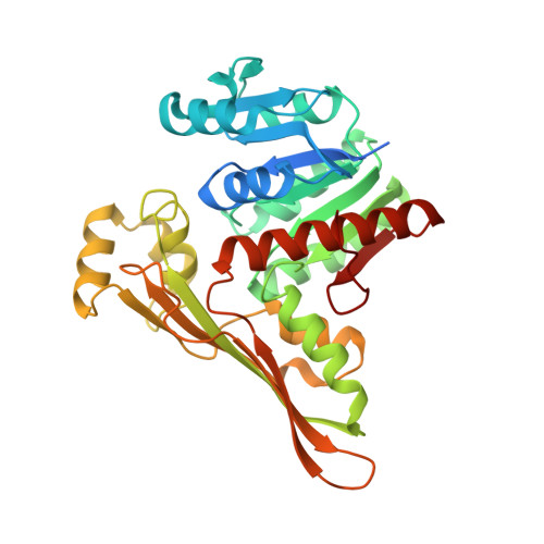
A suicide enzyme catalyzes multiple reactions for biotin biosynthesis in cyanobacteria.

Sakaki, K.Ohishi, K.Shimizu, T.Kobayashi, I.Mori, N.Matsuda, K.Tomita, T.Watanabe, H.Tanaka, K.Kuzuyama, T.Nishiyama, M.

(2020) Nat Chem Biol 16: 415-422

  • DOI: https://doi.org/10.1038/s41589-019-0461-9
  • Primary Citation of Related Structures:  
    6IR4, 6ITD, 6K36, 6K37, 6K38

  • PubMed Abstract: 

    In biotin biosynthesis, the conversion of pimeloyl intermediates to biotin is catalyzed by a universal set of four enzymes: BioF, BioA, BioD and BioB. We found that the gene homologous to bioA, the product of which is involved in the conversion of 8-amino-7-oxononanoate (AON) to 7,8-diaminononanoate (DAN), is missing in the genome of the cyanobacterium Synechocystis sp. PCC 6803. We provide structural and biochemical evidence showing that a novel dehydrogenase, BioU, is involved in biotin biosynthesis and functionally replaces BioA. This enzyme catalyzes three reactions: formation of covalent linkage with AON to yield a BioU-DAN conjugate at the ε-amino group of Lys124 of BioU using NAD(P)H, carboxylation of the conjugate to form BioU-DAN-carbamic acid, and release of DAN-carbamic acid using NAD(P) + . In this biosynthetic pathway, BioU is a suicide enzyme that loses the Lys124 amino group after a single round of reaction.


  • Organizational Affiliation

    Biotechnology Research Center, The University of Tokyo, Bunkyo-ku, Tokyo, Japan.


Macromolecules
Find similar proteins by:  (by identity cutoff)  |  3D Structure
Entity ID: 1
MoleculeChains Sequence LengthOrganismDetailsImage
Slr0355 protein353Synechocystis sp. PCC 6803Mutation(s): 0 
Gene Names: slr0355
EC: 2.6.1.121
UniProt
Find proteins for Q55650 (Synechocystis sp. (strain ATCC 27184 / PCC 6803 / Kazusa))
Explore Q55650 
Go to UniProtKB:  Q55650
Entity Groups  
Sequence Clusters30% Identity50% Identity70% Identity90% Identity95% Identity100% Identity
UniProt GroupQ55650
Sequence Annotations
Expand
  • Reference Sequence
Small Molecules
Ligands 1 Unique
IDChains Name / Formula / InChI Key2D Diagram3D Interactions
CWF (Subject of Investigation/LOI)
Query on CWF

Download Ideal Coordinates CCD File 
B [auth A](8S)-8-azanylnonanoic acid
C9 H19 N O2
VXBHEKXWXPQBQM-QMMMGPOBSA-N
Experimental Data & Validation

Experimental Data

  • Method: X-RAY DIFFRACTION
  • Resolution: 2.30 Å
  • R-Value Free:  0.278 (Depositor), 0.270 (DCC) 
  • R-Value Work:  0.196 (Depositor), 0.200 (DCC) 
  • R-Value Observed: 0.200 (Depositor) 
Space Group: P 31 2 1
Unit Cell:
Length ( Å )Angle ( ˚ )
a = 71.114α = 90
b = 71.114β = 90
c = 98.198γ = 120
Software Package:
Software NamePurpose
REFMACrefinement
HKL-2000data reduction
HKL-2000data scaling
PHASERphasing

Structure Validation

View Full Validation Report


Ligand Structure Quality Assessment 

Created with Raphaël 2.3.0Worse 01 BetterLigand structure goodness of fit to experimental dataBest fitted CWFClick on this verticalbar to view details

Entry History 

Deposition Data

Revision History  (Full details and data files)

  • Version 1.0: 2020-02-26
    Type: Initial release
  • Version 1.1: 2020-04-08
    Changes: Database references
  • Version 1.2: 2023-11-22
    Changes: Data collection, Database references, Refinement description
  • Version 1.3: 2024-11-13
    Changes: Structure summary